1UOP

PROLYL OLIGOPEPTIDASE FROM PORCINE BRAIN, S554A MUTANT WITH BOUND PEPTIDE LIGAND GLY-PHE-GLU-PRO


Experimental Data Snapshot

  • Method: X-RAY DIFFRACTION
  • Resolution: 1.85 Å
  • R-Value Free: 0.194 
  • R-Value Work: 0.156 
  • R-Value Observed: 0.158 

wwPDB Validation   3D Report Full Report


This is version 1.5 of the entry. See complete history


Literature

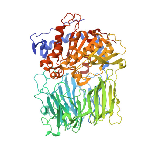
Electrostatic Environment at the Active Site of Prolyl Oligopeptidase is Highly Influential During Substrate Binding

Szeltner, Z.Rea, D.Renner, V.Juliano, L.Fulop, V.Polgar, L.

(2003) J Biol Chem 278: 48786

  • DOI: https://doi.org/10.1074/jbc.M309555200
  • Primary Citation of Related Structures:  
    1UOO, 1UOP, 1UOQ

  • PubMed Abstract: 

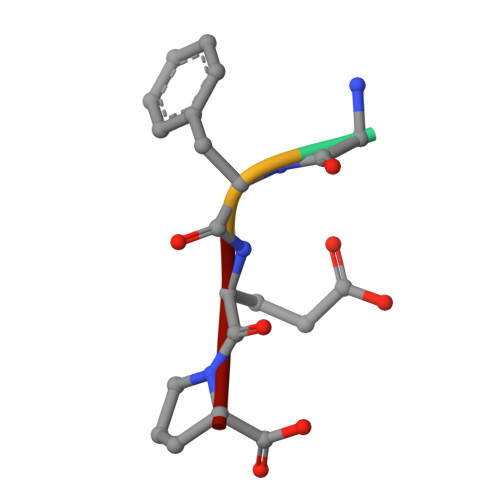
    The positive electrostatic environment of the active site of prolyl oligopeptidase was investigated by using substrates with glutamic acid at positions P2, P3, P4, and P5, respectively. The different substrates gave various pH rate profiles. The pKa values extracted from the curves are apparent parameters, presumably affected by the nearby charged residues, and do not reflect the ionization of a simple catalytic histidine as found in the classic serine peptidases like chymotrypsin and subtilisin. The temperature dependence of kcat/Km did not produce linear Arrhenius plots, indicating different changes in the individual rate constants with the increase in temperature. This rendered it possible to calculate these constants, i.e. the formation (k1) and decomposition (k-1) of the enzyme-substrate complex and the acylation constant (k2), as well as the corresponding activation energies. The results have revealed the relationship between the complex Michaelis parameters and the individual rate constants. Structure determination of the enzyme-substrate complexes has shown that the different substrates display a uniform binding mode. None of the glutamic acids interacts with a charged group. We conclude that the specific rate constant is controlled by k1 rather than k2 and that the charged residues from the substrate and the enzyme can markedly affect the formation but not the structure of the enzyme-substrate complexes.


  • Organizational Affiliation

    Institute of Enzymology, Biological Research Center, Hungarian Academy of Sciences, PO Box 7, H-1518 Budapest 112, Hungary.


Macromolecules
Find similar proteins by:  (by identity cutoff)  |  3D Structure
Entity ID: 1
MoleculeChains Sequence LengthOrganismDetailsImage
PROLYL ENDOPEPTIDASE710Sus scrofaMutation(s): 1 
EC: 3.4.21.26
UniProt
Find proteins for P23687 (Sus scrofa)
Explore P23687 
Go to UniProtKB:  P23687
Entity Groups  
Sequence Clusters30% Identity50% Identity70% Identity90% Identity95% Identity100% Identity
UniProt GroupP23687
Sequence Annotations
Expand
  • Reference Sequence

Find similar proteins by:  Sequence   |   3D Structure  

Entity ID: 2
MoleculeChains Sequence LengthOrganismDetailsImage
PEPTIDE LIGAND GLY-PHE-GLU-PRO4synthetic constructMutation(s): 0 
Sequence Annotations
Expand
  • Reference Sequence
Experimental Data & Validation

Experimental Data

  • Method: X-RAY DIFFRACTION
  • Resolution: 1.85 Å
  • R-Value Free: 0.194 
  • R-Value Work: 0.156 
  • R-Value Observed: 0.158 
  • Space Group: P 21 21 21
Unit Cell:
Length ( Å )Angle ( ˚ )
a = 70.7α = 90
b = 99.3β = 90
c = 110.7γ = 90
Software Package:
Software NamePurpose
REFMACrefinement
DENZOdata reduction
SCALEPACKdata scaling
CCP4phasing

Structure Validation

View Full Validation Report


Entry History 

Deposition Data

Revision History  (Full details and data files)

  • Version 1.0: 2003-10-02
    Type: Initial release
  • Version 1.1: 2011-05-08
    Changes: Version format compliance
  • Version 1.2: 2011-07-13
    Changes: Version format compliance
  • Version 1.3: 2019-07-24
    Changes: Data collection
  • Version 1.4: 2019-10-09
    Changes: Data collection, Other, Source and taxonomy
  • Version 1.5: 2023-12-13
    Changes: Data collection, Database references, Refinement description