1V3O

Crystal structure of d(GCGAGAGC): the DNA quadruplex structure split from the octaplex


Experimental Data Snapshot

  • Method: X-RAY DIFFRACTION
  • Resolution: 1.70 Å
  • R-Value Free: 0.296 
  • R-Value Work: 0.262 

wwPDB Validation   3D Report Full Report


This is version 1.4 of the entry. See complete history


Literature

Crystal structures of a DNA octaplex with I-motif of G-quartets and its splitting into two quadruplexes suggest a folding mechanism of eight tandem repeats

Kondo, J.Adachi, W.Umeda, S.Sunami, T.Takenaka, A.

(2004) Nucleic Acids Res 32: 2541-2549

  • DOI: https://doi.org/10.1093/nar/gkh575
  • Primary Citation of Related Structures:  
    1V3N, 1V3O, 1V3P

  • PubMed Abstract: 

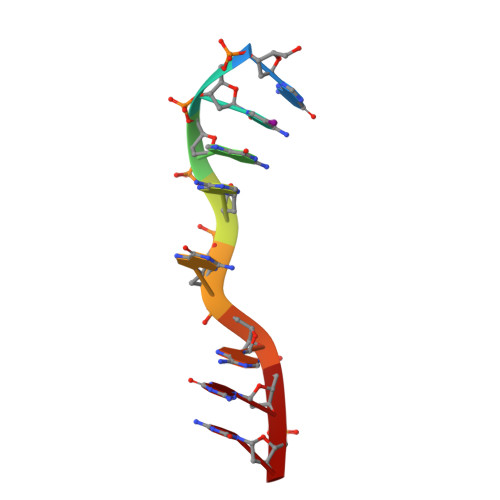
    Recent genomic analyses revealed many kinds of tandem repeats of specific sequences. Some of them are related to genetic diseases, but their biological functions and structures are still unknown. Two X-ray structures of a short DNA fragment d(gcGA[G]1Agc) show that four base-intercalated duplexes are assembled to form an octaplex at a low K+ concentration, in which the eight G5 residues form a stacked double G-quartet in the central part. At a higher K+ concentration, however, the octaplex is split into just two halves. These structural features suggest a folding process of eight tandem repeats of d(ccGA[G]4Agg), according to a double Greek-key motif. Such a packaging of the repeats could facilitate slippage of a certain sequence during DNA replication, to induce increase or decrease of the repeats.


  • Organizational Affiliation

    Graduate School of Bioscience and Biotechnology, Tokyo Institute of Technology, Yokohama 226-8501, Japan.


Macromolecules

Find similar nucleic acids by:  Sequence   |   3D Structure  

Entity ID: 1
MoleculeChains LengthOrganismImage
5'-D(*GP*(C38)P*GP*AP*GP*AP*GP*C)-3'
A, B
8N/A
Sequence Annotations
Expand
  • Reference Sequence
Small Molecules
Ligands 1 Unique
IDChains Name / Formula / InChI Key2D Diagram3D Interactions
K
Query on K

Download Ideal Coordinates CCD File 
C [auth A]POTASSIUM ION
K
NPYPAHLBTDXSSS-UHFFFAOYSA-N
Experimental Data & Validation

Experimental Data

  • Method: X-RAY DIFFRACTION
  • Resolution: 1.70 Å
  • R-Value Free: 0.296 
  • R-Value Work: 0.262 
  • Space Group: I 2 2 2
Unit Cell:
Length ( Å )Angle ( ˚ )
a = 34.65α = 90
b = 42.49β = 90
c = 64.08γ = 90
Software Package:
Software NamePurpose
d*TREKdata scaling
SCALAdata scaling
AMoREphasing
CNSrefinement
d*TREKdata reduction
CCP4data scaling

Structure Validation

View Full Validation Report


Entry History 

Deposition Data

Revision History  (Full details and data files)

  • Version 1.0: 2004-06-08
    Type: Initial release
  • Version 1.1: 2008-04-27
    Changes: Version format compliance
  • Version 1.2: 2011-07-13
    Changes: Version format compliance
  • Version 1.3: 2014-04-16
    Changes: Other
  • Version 1.4: 2023-10-25
    Changes: Data collection, Database references, Derived calculations, Refinement description