


Crystal structure of hypothetical protein (tm1410) from THERMOTOGA MARITIMA at 2.20 A resolution
Joint Center for Structural Genomics (JCSG)To be published.
Experimental Data Snapshot
wwPDB Validation 3D Report Full Report

Entity ID: 1 | |||||
|---|---|---|---|---|---|
| Molecule | Chains | Sequence Length | Organism | Details | Image |
| Hypothetical protein TM1410 | 309 | Thermotoga maritima | Mutation(s): 6 Gene Names: tm1410 | 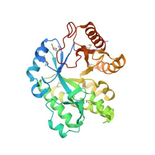 | |
UniProt | |||||
Find proteins for Q9X1D0 (Thermotoga maritima (strain ATCC 43589 / DSM 3109 / JCM 10099 / NBRC 100826 / MSB8)) Explore Q9X1D0 Go to UniProtKB: Q9X1D0 | |||||
Entity Groups | |||||
| Sequence Clusters | 30% Identity50% Identity70% Identity90% Identity95% Identity100% Identity | ||||
| UniProt Group | Q9X1D0 | ||||
Sequence AnnotationsExpand | |||||
| |||||
| Ligands 2 Unique | |||||
|---|---|---|---|---|---|
| ID | Chains | Name / Formula / InChI Key | 2D Diagram | 3D Interactions | |
| GOL Query on GOL | AA [auth C] BA [auth C] CA [auth C] DA [auth C] FA [auth D] | GLYCEROL C3 H8 O3 PEDCQBHIVMGVHV-UHFFFAOYSA-N |  | ||
| UNL Query on UNL | EA [auth D] G [auth A] KA [auth E] O [auth B] PA [auth F] | Unknown ligand PEDCQBHIVMGVHV-UHFFFAOYSA-N | |||
| Modified Residues 1 Unique | |||||
|---|---|---|---|---|---|
| ID | Chains | Type | Formula | 2D Diagram | Parent |
| MSE Query on MSE | A, B, C, D, E A, B, C, D, E, F | L-PEPTIDE LINKING | C5 H11 N O2 Se |  | MET |
| Length ( Å ) | Angle ( ˚ ) |
|---|---|
| a = 194.93 | α = 90 |
| b = 84.62 | β = 119.9 |
| c = 195.01 | γ = 90 |
| Software Name | Purpose |
|---|---|
| REFMAC | refinement |
| XSCALE | data scaling |
| PDB_EXTRACT | data extraction |
| XDS | data reduction |
| SHELX | phasing |
| SHARP | phasing |


