2DV5 | pdb_00002dv5

Crystal structure of Leu65 to Ala mutant of Diphthine synthase


Experimental Data Snapshot

  • Method: X-RAY DIFFRACTION
  • Resolution: 2.20 Å
  • R-Value Free: 
    0.228 (Depositor), 0.226 (DCC) 
  • R-Value Work: 
    0.198 (Depositor), 0.193 (DCC) 
  • R-Value Observed: 
    0.198 (Depositor) 

Starting Model: experimental
View more details

wwPDB Validation   3D Report Full Report


Ligand Structure Quality Assessment 


This is version 1.5 of the entry. See complete history


Literature

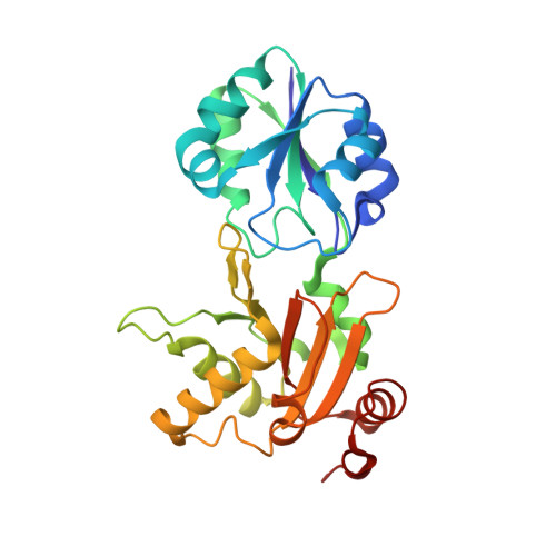
Systematic study on crystal-contact engineering of diphthine synthase: influence of mutations at crystal-packing regions on X-ray diffraction quality.

Mizutani, H.Saraboji, K.Malathy Sony, S.M.Ponnuswamy, M.N.Kumarevel, T.Krishna Swamy, B.S.Simanshu, D.K.Murthy, M.R.Kunishima, N.

(2008) Acta Crystallogr D Biol Crystallogr 64: 1020-1033

  • DOI: https://doi.org/10.1107/S0907444908023019
  • Primary Citation of Related Structures:  
    2DSG, 2DSH, 2DSI, 2DV4, 2DV5, 2DV7, 2DXV, 2DXW, 2DXX, 2E07, 2E08, 2E15, 2E16, 2E17, 2E7R, 2EGB, 2EGL, 2EGS, 2EHC, 2EHL, 2Z6R

  • PubMed Abstract: 

    It is well known that protein crystallizability can be influenced by site-directed mutagenesis of residues on the molecular surface of proteins, indicating that the intermolecular interactions in crystal-packing regions may play a crucial role in the structural regularity at atomic resolution of protein crystals. Here, a systematic examination was made of the improvement in the diffraction resolution of protein crystals on introducing a single mutation of a crystal-packing residue in order to provide more favourable packing interactions, using diphthine synthase from Pyrococcus horikoshii OT3 as a model system. All of a total of 21 designed mutants at 13 different crystal-packing residues yielded almost isomorphous crystals from the same crystallization conditions as those used for the wild-type crystals, which diffracted X-rays to 2.1 A resolution. Of the 21 mutants, eight provided crystals with an improved resolution of 1.8 A or better. Thus, it has been clarified that crystal quality can be improved by introducing a suitable single mutation of a crystal-packing residue. In the improved crystals, more intimate crystal-packing interactions than those in the wild-type crystal are observed. Notably, the mutants K49R and T146R yielded crystals with outstandingly improved resolutions of 1.5 and 1.6 A, respectively, in which a large-scale rearrangement of packing interactions was unexpectedly observed despite the retention of the same isomorphous crystal form. In contrast, the mutants that provided results that were in good agreement with the designed putative structures tended to achieve only moderate improvements in resolution of up to 1.75 A. These results suggest a difficulty in the rational prediction of highly effective mutations in crystal engineering.


  • Organizational Affiliation
    • RIKEN SPring-8 Center, Harima Institute, 1-1-1 Kouto, Sayo-cho, Sayo-gun, Hyogo 679-5148, Japan.

Macromolecules
Find similar proteins by:  (by identity cutoff)  |  3D Structure
Entity ID: 1
MoleculeChains Sequence LengthOrganismDetailsImage
diphthine synthase
A, B
265Pyrococcus horikoshii OT3Mutation(s): 1 
Gene Names: dphB
EC: 2.1.1.98
UniProt
Find proteins for O58456 (Pyrococcus horikoshii (strain ATCC 700860 / DSM 12428 / JCM 9974 / NBRC 100139 / OT-3))
Explore O58456 
Go to UniProtKB:  O58456
Entity Groups  
Sequence Clusters30% Identity50% Identity70% Identity90% Identity95% Identity100% Identity
UniProt GroupO58456
Sequence Annotations
Expand
  • Reference Sequence
Small Molecules
Ligands 4 Unique
IDChains Name / Formula / InChI Key2D Diagram3D Interactions
SAH
Query on SAH

Download Ideal Coordinates CCD File 
F [auth A]S-ADENOSYL-L-HOMOCYSTEINE
C14 H20 N6 O5 S
ZJUKTBDSGOFHSH-WFMPWKQPSA-N
MES
Query on MES

Download Ideal Coordinates CCD File 
G [auth A],
M [auth B]
2-(N-MORPHOLINO)-ETHANESULFONIC ACID
C6 H13 N O4 S
SXGZJKUKBWWHRA-UHFFFAOYSA-N
SO4
Query on SO4

Download Ideal Coordinates CCD File 
C [auth A]
D [auth A]
E [auth A]
I [auth B]
J [auth B]
C [auth A],
D [auth A],
E [auth A],
I [auth B],
J [auth B],
K [auth B],
L [auth B]
SULFATE ION
O4 S
QAOWNCQODCNURD-UHFFFAOYSA-L
GOL
Query on GOL

Download Ideal Coordinates CCD File 
H [auth A],
N [auth B],
O [auth B]
GLYCEROL
C3 H8 O3
PEDCQBHIVMGVHV-UHFFFAOYSA-N
Experimental Data & Validation

Experimental Data

  • Method: X-RAY DIFFRACTION
  • Resolution: 2.20 Å
  • R-Value Free:  0.228 (Depositor), 0.226 (DCC) 
  • R-Value Work:  0.198 (Depositor), 0.193 (DCC) 
  • R-Value Observed: 0.198 (Depositor) 
Space Group: P 41 21 2
Unit Cell:
Length ( Å )Angle ( ˚ )
a = 105.219α = 90
b = 105.219β = 90
c = 137.334γ = 90
Software Package:
Software NamePurpose
CNSrefinement
HKL-2000data reduction
HKL-2000data scaling

Structure Validation

View Full Validation Report


Ligand Structure Quality Assessment 


Entry History 

Deposition Data

Revision History  (Full details and data files)

  • Version 1.0: 2007-01-28
    Type: Initial release
  • Version 1.1: 2008-04-30
    Changes: Version format compliance
  • Version 1.2: 2011-07-13
    Changes: Source and taxonomy, Version format compliance
  • Version 1.3: 2021-11-10
    Changes: Database references, Derived calculations
  • Version 1.4: 2023-10-25
    Changes: Data collection, Refinement description
  • Version 1.5: 2025-08-06
    Changes: Data collection, Database references, Structure summary