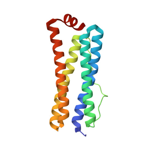
2FL0

Oxidized (All ferric) form of the Azotobacter vinelandii bacterioferritin


Experimental Data Snapshot

  • Method: X-RAY DIFFRACTION
  • Resolution: 2.70 Å
  • R-Value Free: 0.256 
  • R-Value Work: 0.203 
  • R-Value Observed: 0.203 

wwPDB Validation   3D Report Full Report


Ligand Structure Quality Assessment 


This is version 2.0 of the entry. See complete history


Literature

Redox-Dependent Structural Changes in the Azotobacter vinelandii Bacterioferritin: New Insights into the Ferroxidase and Iron Transport Mechanism(,).

Swartz, L.Kuchinskas, M.Li, H.Poulos, T.L.Lanzilotta, W.N.

(2006) Biochemistry 45: 4421-4428

  • DOI: https://doi.org/10.1021/bi060146w
  • Primary Citation of Related Structures:  
    2FKZ, 2FL0

  • PubMed Abstract: 

    In this work, we report the X-ray crystal structure of the aerobically isolated (oxidized) and the anaerobic dithionite-reduced (at pH 8.0) forms of the native Azotobacter vinelandii bacterioferritin to 2.7 and 2.0 A resolution, respectively. Iron K-edge multiple anomalous dispersion (MAD) experiments unequivocally identified the presence of three independent iron-containing sites within the protein structure. Specifically, a dinuclear (ferroxidase) site, a b-type heme site, and the binding of a single iron atom at the four-fold molecular axis of the protein shell were observed. In addition to the novel observation of iron at the four-fold pore, these data also reveal that the oxidized form of the protein has a symmetrical ferroxidase site containing two five-coordinate iron atoms. Each iron atom is ligated by four carboxylate oxygen atoms and a single histidyl nitrogen atom. A single water molecule is found within hydrogen bonding distance of the ferroxidase site that bridges the two iron atoms on the side opposite the histidine ligands. Chemical reduction of the protein under anaerobic conditions results in an increase in the average Fe-Fe distance in the ferroxidase site from approximately 3.5 to approximately 4.0 A and the loss of one of the ligands, H130. In addition, there is significant movement of the bridging water molecule and several other amino acid side chains in the vicinity of the ferroxidase site and along the D helix to the three-fold symmetry axis. In contrast to previous work, the higher-resolution data for the dithionite-reduced structure suggest that the heme may be bound in multiple conformations. Taken together, these data allow a molecular movie of the ferroxidase gating mechanism to be developed and provide further insight into the iron uptake and/or release and mineralization mechanism of bacterioferritins in general.


  • Organizational Affiliation

    Department of Molecular Biology and Biochemistry, University of California, Irvine, California 92697, USA.


Macromolecules
Find similar proteins by:  (by identity cutoff)  |  3D Structure
Entity ID: 1
MoleculeChains Sequence LengthOrganismDetailsImage
Bacterioferritin
A, B, C, D, E
A, B, C, D, E, F, G, H
155Azotobacter vinelandiiMutation(s): 0 
EC: 1.16.3.1
UniProt
Find proteins for P22759 (Azotobacter vinelandii)
Explore P22759 
Go to UniProtKB:  P22759
Entity Groups  
Sequence Clusters30% Identity50% Identity70% Identity90% Identity95% Identity100% Identity
UniProt GroupP22759
Sequence Annotations
Expand
  • Reference Sequence
Small Molecules
Ligands 3 Unique
IDChains Name / Formula / InChI Key2D Diagram3D Interactions
HEM
Query on HEM

Download Ideal Coordinates CCD File 
FA [auth E],
NA [auth G],
O [auth A],
X [auth C]
PROTOPORPHYRIN IX CONTAINING FE
C34 H32 Fe N4 O4
KABFMIBPWCXCRK-RGGAHWMASA-L
FE2
Query on FE2

Download Ideal Coordinates CCD File 
DA [auth E]
EA [auth E]
GA [auth F]
HA [auth F]
I [auth A]
DA [auth E],
EA [auth E],
GA [auth F],
HA [auth F],
I [auth A],
J [auth A],
JA [auth G],
K [auth A],
KA [auth G],
OA [auth H],
P [auth B],
PA [auth H],
Q [auth B],
R [auth B],
U [auth C],
V [auth C],
Y [auth D],
Z [auth D]
FE (II) ION
Fe
CWYNVVGOOAEACU-UHFFFAOYSA-N
MG
Query on MG

Download Ideal Coordinates CCD File 
AA [auth D]
BA [auth D]
CA [auth D]
IA [auth F]
L [auth A]
AA [auth D],
BA [auth D],
CA [auth D],
IA [auth F],
L [auth A],
LA [auth G],
M [auth A],
MA [auth G],
N [auth A],
QA [auth H],
S [auth B],
T [auth B],
W [auth C]
MAGNESIUM ION
Mg
JLVVSXFLKOJNIY-UHFFFAOYSA-N
Experimental Data & Validation

Experimental Data

  • Method: X-RAY DIFFRACTION
  • Resolution: 2.70 Å
  • R-Value Free: 0.256 
  • R-Value Work: 0.203 
  • R-Value Observed: 0.203 
  • Space Group: H 3
Unit Cell:
Length ( Å )Angle ( ˚ )
a = 124.48α = 90
b = 124.48β = 90
c = 285.112γ = 120
Software Package:
Software NamePurpose
CNSrefinement
HKL-2000data reduction
SCALEPACKdata scaling
SHARPphasing

Structure Validation

View Full Validation Report


Ligand Structure Quality Assessment 


Entry History 

Deposition Data

Revision History  (Full details and data files)

  • Version 1.0: 2006-04-04
    Type: Initial release
  • Version 1.1: 2008-05-01
    Changes: Version format compliance
  • Version 1.2: 2011-07-13
    Changes: Derived calculations, Version format compliance
  • Version 2.0: 2024-02-14
    Changes: Atomic model, Data collection, Database references, Derived calculations