2OQA | pdb_00002oqa

X-ray Sequence and Crystal Structure of Luffaculin 1, a Novel Type 1 Ribosome-inactivating Protein


Experimental Data Snapshot

  • Method: X-RAY DIFFRACTION
  • Resolution: 1.40 Å
  • R-Value Free: 
    0.232 (Depositor) 
  • R-Value Work: 
    0.213 (Depositor), 0.215 (DCC) 
  • R-Value Observed: 
    0.213 (Depositor) 

Starting Model: in silico
View more details

wwPDB Validation   3D Report Full Report


Ligand Structure Quality Assessment 


This is version 1.6 of the entry. See complete history


Literature

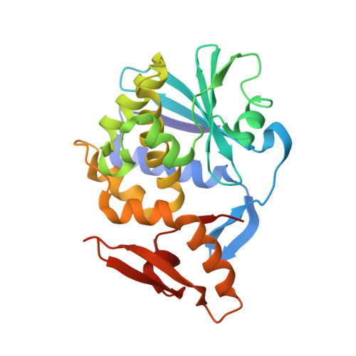
X-ray sequence and crystal structure of luffaculin 1, a novel type 1 ribosome-inactivating protein

Hou, X.Chen, M.Chen, L.Meehan, E.J.Xie, J.Huang, M.

(2007) BMC Struct Biol 7: 29-29

  • DOI: https://doi.org/10.1186/1472-6807-7-29
  • Primary Citation of Related Structures:  
    2OQA

  • PubMed Abstract: 

    Protein sequence can be obtained through Edman degradation, mass spectrometry, or cDNA sequencing. High resolution X-ray crystallography can also be used to derive protein sequence information, but faces the difficulty in distinguishing the Asp/Asn, Glu/Gln, and Val/Thr pairs. Luffaculin 1 is a new type 1 ribosome-inactivating protein (RIP) isolated from the seeds of Luffa acutangula. Besides rRNA N-glycosidase activity, luffaculin 1 also demonstrates activities including inhibiting tumor cells' proliferation and inducing tumor cells' differentiation. The crystal structure of luffaculin 1 was determined at 1.4 A resolution. Its amino-acid sequence was derived from this high resolution structure using the following criteria: 1) high resolution electron density; 2) comparison of electron density between two molecules that exist in the same crystal; 3) evaluation of the chemical environment of residues to break down the sequence assignment ambiguity in residue pairs Glu/Gln, Asp/Asn, and Val/Thr; 4) comparison with sequences of the homologous proteins. Using the criteria 1 and 2, 66% of the residues can be assigned. By incorporating with criterion 3, 86% of the residues were assigned, suggesting the effectiveness of chemical environment evaluation in breaking down residue ambiguity. In total, 94% of the luffaculin 1 sequence was assigned with high confidence using this improved X-ray sequencing strategy. Two N-acetylglucosamine moieties, linked respectively to the residues Asn77 and Asn84, can be identified in the structure. Residues Tyr70, Tyr110, Glu159 and Arg162 define the active site of luffaculin 1 as an RNA N-glycosidase. X-ray sequencing method can be effective to derive sequence information of proteins. The evaluation of the chemical environment of residues is a useful method to break down the assignment ambiguity in Glu/Gln, Asp/Asn, and Val/Thr pairs. The sequence and the crystal structure confirm that luffaculin 1 is a new type 1 RIP.


  • Organizational Affiliation
    • State Key Laboratory of Structural Chemistry, Fujian Institute of Research on the Structure of Matter, The Chinese Academy of Sciences, 155 Yang Qiao Xi Lu, Fuzhou, Fujian, China. houxiaomin@fjirsm.ac.cn <houxiaomin@fjirsm.ac.cn>

Macromolecules
Find similar proteins by:  (by identity cutoff)  |  3D Structure
Entity ID: 1
MoleculeChains Sequence LengthOrganismDetailsImage
Luffaculin 1
A, B
241Luffa acutangulaMutation(s): 0 
EC: 3.2.2.22
UniProt
Find proteins for Q00465 (Luffa aegyptiaca)
Explore Q00465 
Go to UniProtKB:  Q00465
Entity Groups  
Sequence Clusters30% Identity50% Identity70% Identity90% Identity95% Identity100% Identity
UniProt GroupQ00465
Sequence Annotations
Expand
  • Reference Sequence
Small Molecules
Ligands 3 Unique
IDChains Name / Formula / InChI Key2D Diagram3D Interactions
NAG
Query on NAG

Download Ideal Coordinates CCD File 
C [auth A],
D [auth A],
F [auth B],
G [auth B]
2-acetamido-2-deoxy-beta-D-glucopyranose
C8 H15 N O6
OVRNDRQMDRJTHS-FMDGEEDCSA-N
PG4
Query on PG4

Download Ideal Coordinates CCD File 
E [auth A]TETRAETHYLENE GLYCOL
C8 H18 O5
UWHCKJMYHZGTIT-UHFFFAOYSA-N
PEG
Query on PEG

Download Ideal Coordinates CCD File 
H [auth B],
I [auth B]
DI(HYDROXYETHYL)ETHER
C4 H10 O3
MTHSVFCYNBDYFN-UHFFFAOYSA-N
Experimental Data & Validation

Experimental Data

  • Method: X-RAY DIFFRACTION
  • Resolution: 1.40 Å
  • R-Value Free:  0.232 (Depositor) 
  • R-Value Work:  0.213 (Depositor), 0.215 (DCC) 
  • R-Value Observed: 0.213 (Depositor) 
Space Group: P 1
Unit Cell:
Length ( Å )Angle ( ˚ )
a = 39.135α = 89.07
b = 46.813β = 80.01
c = 83.571γ = 72.14
Software Package:
Software NamePurpose
REFMACrefinement
HKL-2000data collection
HKL-2000data reduction
SCALEPACKdata scaling
AMoREphasing

Structure Validation

View Full Validation Report


Ligand Structure Quality Assessment 


Entry History 

Deposition Data

Revision History  (Full details and data files)

  • Version 1.0: 2007-05-29
    Type: Initial release
  • Version 1.1: 2008-05-01
    Changes: Version format compliance
  • Version 1.2: 2011-07-13
    Changes: Non-polymer description, Version format compliance
  • Version 1.3: 2020-07-29
    Type: Remediation
    Reason: Carbohydrate remediation
    Changes: Data collection, Derived calculations, Structure summary
  • Version 1.4: 2023-12-27
    Changes: Data collection, Database references, Structure summary
  • Version 1.5: 2024-04-03
    Changes: Refinement description
  • Version 1.6: 2024-12-25
    Changes: Advisory, Derived calculations, Structure summary