2OVV | pdb_00002ovv

Crystal structure of the catalytic domain of rat phosphodiesterase 10A


Experimental Data Snapshot

  • Method: X-RAY DIFFRACTION
  • Resolution: 2.00 Å
  • R-Value Free: 
    0.206 (Depositor), 0.214 (DCC) 
  • R-Value Work: 
    0.174 (Depositor), 0.185 (DCC) 
  • R-Value Observed: 
    0.177 (Depositor) 

wwPDB Validation   3D Report Full Report


Ligand Structure Quality Assessment 


This is version 1.3 of the entry. See complete history


Literature

Discovery of a series of 6,7-dimethoxy-4-pyrrolidylquinazoline PDE10A inhibitors

Chappie, T.A.Humphrey, J.M.Allen, M.P.Estep, K.G.Fox, C.B.Lebel, L.A.Liras, S.Marr, E.S.Menniti, F.S.Pandit, J.Schmidt, C.J.Tu, M.Williams, R.D.Yang, F.V.

(2007) J Med Chem 50: 182-185

  • DOI: https://doi.org/10.1021/jm060653b
  • Primary Citation of Related Structures:  
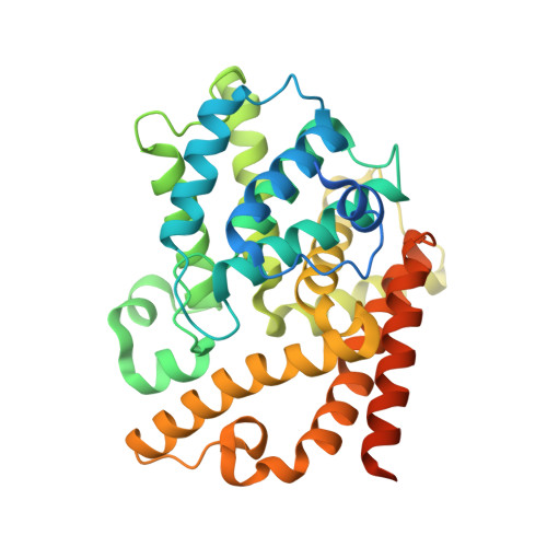
    2O8H, 2OVV, 2OVY

  • PubMed Abstract: 

    A papaverine based pharmacophore model for PDE10A inhibition was generated via SBDD and used to design a library of 4-amino-6,7-dimethoxyquinazolines. From this library emerged an aryl ether pyrrolidyl 6,7-dimethoxyquinazoline series that became the focal point for additional modeling, X-ray, and synthetic efforts toward increasing PDE10A inhibitory potency and selectivity versus PDE3A/B. These efforts culminated in the discovery of 29, a potent and selective brain penetrable inhibitor of PDE10A.


  • Organizational Affiliation
    • CNS Discovery and Experimental Medicinal Sciences, Pfizer Global Research and Development, Eastern Point Road, Groton, Connecticut 06340, USA. thomas.a.chappie@pfizer.com

Macromolecules
Find similar proteins by:  (by identity cutoff)  |  3D Structure
Entity ID: 1
MoleculeChains Sequence LengthOrganismDetailsImage
Phosphodiesterase-10A362Rattus norvegicusMutation(s): 0 
Gene Names: Pde10aPDE10A
EC: 3.1.4.17
UniProt
Find proteins for Q9QYJ6 (Rattus norvegicus)
Explore Q9QYJ6 
Go to UniProtKB:  Q9QYJ6
Entity Groups  
Sequence Clusters30% Identity50% Identity70% Identity90% Identity95% Identity100% Identity
UniProt GroupQ9QYJ6
Sequence Annotations
Expand
  • Reference Sequence
Experimental Data & Validation

Experimental Data

  • Method: X-RAY DIFFRACTION
  • Resolution: 2.00 Å
  • R-Value Free:  0.206 (Depositor), 0.214 (DCC) 
  • R-Value Work:  0.174 (Depositor), 0.185 (DCC) 
  • R-Value Observed: 0.177 (Depositor) 
Space Group: H 3
Unit Cell:
Length ( Å )Angle ( ˚ )
a = 120.657α = 90
b = 120.657β = 90
c = 83.273γ = 120
Software Package:
Software NamePurpose
d*TREKdata scaling
REFMACrefinement
PDB_EXTRACTdata extraction

Structure Validation

View Full Validation Report


Ligand Structure Quality Assessment 


Entry History 

Deposition Data

Revision History  (Full details and data files)

  • Version 1.0: 2007-03-06
    Type: Initial release
  • Version 1.1: 2008-05-01
    Changes: Version format compliance
  • Version 1.2: 2011-07-13
    Changes: Derived calculations, Version format compliance
  • Version 1.3: 2024-12-25
    Changes: Advisory, Data collection, Database references, Derived calculations, Structure summary