2Q1M | pdb_00002q1m

Crystal Structure of human GITRL


Experimental Data Snapshot

  • Method: X-RAY DIFFRACTION
  • Resolution: 2.30 Å
  • R-Value Free: 
    0.257 (Depositor), 0.243 (DCC) 
  • R-Value Work: 
    0.215 (Depositor), 0.209 (DCC) 
  • R-Value Observed: 
    0.217 (Depositor) 

wwPDB Validation   3D Report Full Report


This is version 1.3 of the entry. See complete history


Literature

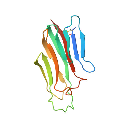
Assembly and structural properties of glucocorticoid-induced TNF receptor ligand: Implications for function.

Chattopadhyay, K.Ramagopal, U.A.Mukhopadhaya, A.Malashkevich, V.N.Dilorenzo, T.P.Brenowitz, M.Nathenson, S.G.Almo, S.C.

(2007) Proc Natl Acad Sci U S A 104: 19452-19457

  • DOI: https://doi.org/10.1073/pnas.0709264104
  • Primary Citation of Related Structures:  
    2Q1M, 2R30, 2R32

  • PubMed Abstract: 

    Glucocorticoid-induced TNF receptor ligand (GITRL), a recently identified member of the TNF family, binds to its receptor GITR on both effector and regulatory T cells and generates positive costimulatory signals implicated in a wide range of T cell functions. Structural analysis reveals that the human GITRL (hGITRL) ectodomain self-assembles into an atypical expanded homotrimer with sparse monomer-monomer interfaces. Consistent with the small intersubunit interfaces, hGITRL exhibits a relatively weak tendency to trimerize in solution and displays a monomer-trimer equilibrium not reported for other TNF family members. This unique assembly behavior has direct implications for hGITRL-GITR signaling, because enforced trimerization of soluble hGITRL ectodomain results in an approximately 100-fold increase in its receptor binding affinity and also in enhanced costimulatory activity. The apparent reduction in affinity that is the consequence of this dynamic equilibrium may represent a mechanism to realize the biologically optimal level of signaling through the hGITRL-GITR pathway, as opposed to the maximal achievable level.


  • Organizational Affiliation
    • Departments of Microbiology and Immunology, Cell Biology, Biochemistry, Physiology and Biophysics, Medicine (Division of Endocrinology), Albert Einstein College of Medicine, Bronx, NY 10461, USA.

Macromolecules
Find similar proteins by:  (by identity cutoff)  |  3D Structure
Entity ID: 1
MoleculeChains Sequence LengthOrganismDetailsImage
Tumor necrosis factor ligand superfamily member 18130Homo sapiensMutation(s): 0 
Gene Names: TNFSF18AITRLGITRLTL6
UniProt & NIH Common Fund Data Resources
Find proteins for Q9UNG2 (Homo sapiens)
Explore Q9UNG2 
Go to UniProtKB:  Q9UNG2
PHAROS:  Q9UNG2
GTEx:  ENSG00000120337 
Entity Groups  
Sequence Clusters30% Identity50% Identity70% Identity90% Identity95% Identity100% Identity
UniProt GroupQ9UNG2
Sequence Annotations
Expand
  • Reference Sequence
Experimental Data & Validation

Experimental Data

  • Method: X-RAY DIFFRACTION
  • Resolution: 2.30 Å
  • R-Value Free:  0.257 (Depositor), 0.243 (DCC) 
  • R-Value Work:  0.215 (Depositor), 0.209 (DCC) 
  • R-Value Observed: 0.217 (Depositor) 
Space Group: P 63
Unit Cell:
Length ( Å )Angle ( ˚ )
a = 72.691α = 90
b = 72.691β = 90
c = 53.101γ = 120
Software Package:
Software NamePurpose
DENZOdata reduction
SCALEPACKdata scaling
REFMACrefinement
PDB_EXTRACTdata extraction
ADSCdata collection
HKL-2000data reduction
SHELXDphasing
SHELXEmodel building

Structure Validation

View Full Validation Report


Entry History 

Deposition Data

Revision History  (Full details and data files)

  • Version 1.0: 2007-11-13
    Type: Initial release
  • Version 1.1: 2011-07-13
    Changes: Version format compliance
  • Version 1.2: 2017-10-18
    Changes: Refinement description
  • Version 1.3: 2024-10-16
    Changes: Data collection, Database references, Structure summary