3CVV

Drosophila melanogaster (6-4) photolyase bound to ds DNA with a T-T (6-4) photolesion and F0 cofactor


Experimental Data Snapshot

  • Method: X-RAY DIFFRACTION
  • Resolution: 2.10 Å
  • R-Value Free: 0.213 
  • R-Value Work: 0.166 
  • R-Value Observed: 0.168 

wwPDB Validation   3D Report Full Report


Ligand Structure Quality Assessment 


This is version 1.3 of the entry. See complete history


Literature

The archaeal cofactor F0 is a light-harvesting antenna chromophore in eukaryotes.

Glas, A.F.Maul, M.J.Cryle, M.Barends, T.R.Schneider, S.Kaya, E.Schlichting, I.Carell, T.

(2009) Proc Natl Acad Sci U S A 106: 11540-11545

  • DOI: https://doi.org/10.1073/pnas.0812665106
  • Primary Citation of Related Structures:  
    3CVV

  • PubMed Abstract: 

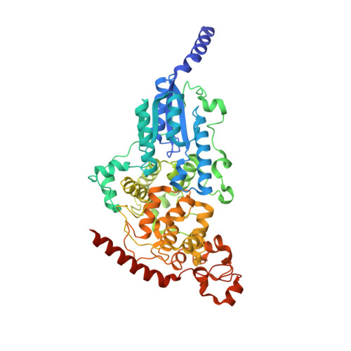
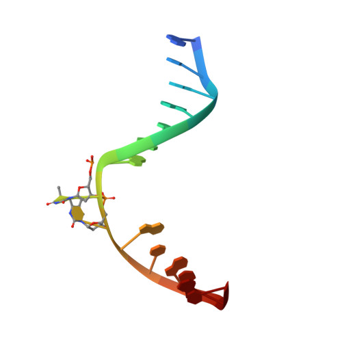
    Archae possess unique biochemical systems quite distinct from the pathways present in eukaryotes and eubacteria. 7,8-Dimethyl-8-hydroxy-5deazaflavin (F(0)) and F(420) are unique deazaflavin-containing coenzyme and methanogenic signature molecules, essential for a variety of biochemical transformations associated with methane biosynthesis and light-dependent DNA repair. The deazaflavin cofactor system functions during methane biosynthesis as a low-potential hydrid shuttle F(420)/F(420)H(2). In DNA photolyase repair proteins, the deazaflavin cofactor is in the deprotonated state active as a light-collecting energy transfer pigment. As such, it converts blue sunlight into energy used by the proteins to drive an essential repair process. Analysis of a eukaryotic (6-4) DNA photolyase from Drosophila melanogaster revealed a binding pocket, which tightly binds F(0). Residues in the pocket activate the cofactor by deprotonation so that light absorption and energy transfer are switched on. The crystal structure of F(0) in complex with the D. melanogaster protein shows the atomic details of F(0) binding and activation, allowing characterization of the residues involved in F(0) activation. The results show that the F(0)/F(420) coenzyme system, so far believed to be strictly limited to the archael kingdom of life, is far more widespread than anticipated. Analysis of a D. melanogaster extract and of a DNA photolyase from the primitive eukaryote Ostreococcus tauri provided direct proof for the presence of the F(0) cofactor also in higher eukaryotes.


  • Organizational Affiliation

    Department of Chemistry and Biochemistry, Ludwig-Maximilians University Munich, Centre for Integrative Protein Science, 81377 Munich, Germany.


Macromolecules

Find similar proteins by:  (by identity cutoff)  |  3D Structure
Entity ID: 3
MoleculeChains Sequence LengthOrganismDetailsImage
RE11660pC [auth A]543Drosophila melanogasterMutation(s): 0 
Gene Names: phr64-520
UniProt
Find proteins for Q8SXK5 (Drosophila melanogaster)
Explore Q8SXK5 
Go to UniProtKB:  Q8SXK5
Entity Groups  
Sequence Clusters30% Identity50% Identity70% Identity90% Identity95% Identity100% Identity
UniProt GroupQ8SXK5
Sequence Annotations
Expand
  • Reference Sequence

Find similar nucleic acids by:  Sequence   |   3D Structure  

Entity ID: 1
MoleculeChains LengthOrganismImage
DNA (5'-D(*DAP*DCP*DAP*DGP*DCP*DGP*DGP*(64T)P*(5PY)P*DGP*DCP*DAP*DGP*DGP*DT)-3')A [auth C]15N/A
Sequence Annotations
Expand
  • Reference Sequence

Find similar nucleic acids by:  Sequence   |   3D Structure  

Entity ID: 2
MoleculeChains LengthOrganismImage
DNA (5'-D(*DTP*DAP*DCP*DCP*DTP*DGP*DCP*DAP*DAP*DCP*DCP*DGP*DCP*DTP*DG)-3')B [auth D]15N/A
Sequence Annotations
Expand
  • Reference Sequence
Experimental Data & Validation

Experimental Data

  • Method: X-RAY DIFFRACTION
  • Resolution: 2.10 Å
  • R-Value Free: 0.213 
  • R-Value Work: 0.166 
  • R-Value Observed: 0.168 
  • Space Group: P 21 21 21
Unit Cell:
Length ( Å )Angle ( ˚ )
a = 84.59α = 90
b = 91.07β = 90
c = 88.26γ = 90
Software Package:
Software NamePurpose
REFMACrefinement
MAR345data collection
XDSdata reduction
XSCALEdata scaling
REFMACphasing

Structure Validation

View Full Validation Report


Ligand Structure Quality Assessment 


Entry History 

Deposition Data

Revision History  (Full details and data files)

  • Version 1.0: 2009-06-16
    Type: Initial release
  • Version 1.1: 2011-07-13
    Changes: Version format compliance
  • Version 1.2: 2017-10-25
    Changes: Refinement description
  • Version 1.3: 2024-02-21
    Changes: Data collection, Database references, Derived calculations