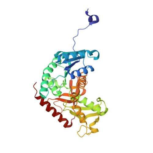
3H3F

Rabbit muscle L-lactate dehydrogenase in complex with NADH and oxamate


Experimental Data Snapshot

  • Method: X-RAY DIFFRACTION
  • Resolution: 2.38 Å
  • R-Value Free: 0.207 
  • R-Value Work: 0.160 
  • R-Value Observed: 0.162 

Starting Model: experimental
View more details

wwPDB Validation   3D Report Full Report


Ligand Structure Quality Assessment 


This is version 1.3 of the entry. See complete history


Literature

Modeling of isotope effects on binding oxamate to lactic dehydrogenase

Swiderek, K.Panczakiewicz, A.Bujacz, A.Bujacz, G.Paneth, P.

(2009) J Phys Chem B 113: 12782-12789

  • DOI: https://doi.org/10.1021/jp903579x
  • Primary Citation of Related Structures:  
    3H3F

  • PubMed Abstract: 

    A new crystal structure of the rabbit muscle L-lactic dehydrogenase (PDB code 3H3F) has been determined. The independent unit of this structure contains two tetramers, each of them with a unique constitution of two active sites with the open loop conformation and two with the loops closed over the actives sites. On the basis of this structure, interactions of an inhibitor, oxamate anion, with the protein have been modeled using different hybrid schemes that involved B3LYP/6-31++G(d,p) DFT theory level in the QM layer. In ONIOM calculations, either Amber (QM:MM) or one of the three semiempirical parametrizations, AM1, PM3, and RM1 (QM:QM) was used, while in the traditional QM/MM scheme, the OPLS-AA force field was used for the outer layer. Normal modes of vibrations of oxamate in aqueous solution and in the active site of the enzyme were used to calculate binding isotope effects. On the basis of the comparison of the values obtained theoretically with those experimentally determined for the oxygen atoms of the carboxylic group of oxamate it was concluded that the DFT/OPLS-AA scheme, applied to the dimer consisting of two chains, one with the open loop and the other with the closed loop conformation, provides the best description of the active site. Calculations of the binding isotope effects of the other atoms of oxamate suggest that nitrogen isotope effect may be useful for the experimental differentiation between open and closed loop conformations.


  • Organizational Affiliation

    Institute of Applied Radiation Chemistry, Technical University of Lodz, ul. Zeromskiego 116, 90-924 Lodz, Poland.


Macromolecules
Find similar proteins by:  (by identity cutoff)  |  3D Structure
Entity ID: 1
MoleculeChains Sequence LengthOrganismDetailsImage
L-lactate dehydrogenase A chain
A, B, C, D, E
A, B, C, D, E, F, G, H
331Oryctolagus cuniculusMutation(s): 0 
EC: 1.1.1.27
UniProt
Find proteins for P13491 (Oryctolagus cuniculus)
Explore P13491 
Go to UniProtKB:  P13491
Entity Groups  
Sequence Clusters30% Identity50% Identity70% Identity90% Identity95% Identity100% Identity
UniProt GroupP13491
Sequence Annotations
Expand
  • Reference Sequence
Small Molecules
Ligands 3 Unique
IDChains Name / Formula / InChI Key2D Diagram3D Interactions
NAI
Query on NAI

Download Ideal Coordinates CCD File 
GA [auth F]
I [auth A]
KA [auth G]
L [auth B]
O [auth C]
GA [auth F],
I [auth A],
KA [auth G],
L [auth B],
O [auth C],
PA [auth H],
S [auth D],
Z [auth E]
1,4-DIHYDRONICOTINAMIDE ADENINE DINUCLEOTIDE
C21 H29 N7 O14 P2
BOPGDPNILDQYTO-NNYOXOHSSA-N
OXM
Query on OXM

Download Ideal Coordinates CCD File 
AA [auth E]
HA [auth F]
J [auth A]
LA [auth G]
M [auth B]
AA [auth E],
HA [auth F],
J [auth A],
LA [auth G],
M [auth B],
P [auth C],
QA [auth H],
T [auth D]
OXAMIC ACID
C2 H3 N O3
SOWBFZRMHSNYGE-UHFFFAOYSA-N
ACT
Query on ACT

Download Ideal Coordinates CCD File 
BA [auth E]
CA [auth E]
DA [auth E]
EA [auth E]
FA [auth E]
BA [auth E],
CA [auth E],
DA [auth E],
EA [auth E],
FA [auth E],
IA [auth F],
JA [auth F],
K [auth A],
MA [auth G],
N [auth B],
NA [auth G],
OA [auth G],
Q [auth C],
R [auth C],
RA [auth H],
SA [auth H],
TA [auth H],
U [auth D],
UA [auth H],
V [auth D],
W [auth D],
X [auth D],
Y [auth D]
ACETATE ION
C2 H3 O2
QTBSBXVTEAMEQO-UHFFFAOYSA-M
Binding Affinity Annotations 
IDSourceBinding Affinity
OXM BindingDB:  3H3F Ki: min: 2.60e+4, max: 1.38e+5 (nM) from 3 assay(s)
IC50: min: 5.72e+4, max: 8.00e+4 (nM) from 2 assay(s)
Experimental Data & Validation

Experimental Data

  • Method: X-RAY DIFFRACTION
  • Resolution: 2.38 Å
  • R-Value Free: 0.207 
  • R-Value Work: 0.160 
  • R-Value Observed: 0.162 
  • Space Group: P 1
Unit Cell:
Length ( Å )Angle ( ˚ )
a = 65.495α = 98.49
b = 85.279β = 91.67
c = 138.526γ = 111.59
Software Package:
Software NamePurpose
HKL-2000data collection
MOLREPphasing
REFMACrefinement
DENZOdata reduction
SCALEPACKdata scaling

Structure Validation

View Full Validation Report


Ligand Structure Quality Assessment 


Entry History 

Deposition Data

Revision History  (Full details and data files)

  • Version 1.0: 2009-09-15
    Type: Initial release
  • Version 1.1: 2011-07-13
    Changes: Advisory, Refinement description, Version format compliance
  • Version 1.2: 2014-11-12
    Changes: Structure summary
  • Version 1.3: 2023-11-01
    Changes: Data collection, Database references, Derived calculations, Refinement description