3NOK

Crystal structure of Myxococcus xanthus Glutaminyl Cyclase


Experimental Data Snapshot

  • Method: X-RAY DIFFRACTION
  • Resolution: 1.65 Å
  • R-Value Free: 0.236 
  • R-Value Work: 0.195 
  • R-Value Observed: 0.197 

wwPDB Validation   3D Report Full Report


Ligand Structure Quality Assessment 


This is version 1.4 of the entry. See complete history


Literature

Kinetic and structural characterization of bacterial glutaminyl cyclases from Zymomonas mobilis and Myxococcus xanthus

Carrillo, D.R.Parthier, C.Janckel, N.Grandke, J.Stelter, M.Schilling, S.Boehme, M.Neumann, P.Wolf, R.Demuth, H.U.Stubbs, M.T.Rahfeld, J.U.

(2010) Biol Chem 391: 1419-1428

  • DOI: https://doi.org/10.1515/BC.2010.130
  • Primary Citation of Related Structures:  
    3NOK, 3NOL, 3NOM

  • PubMed Abstract: 

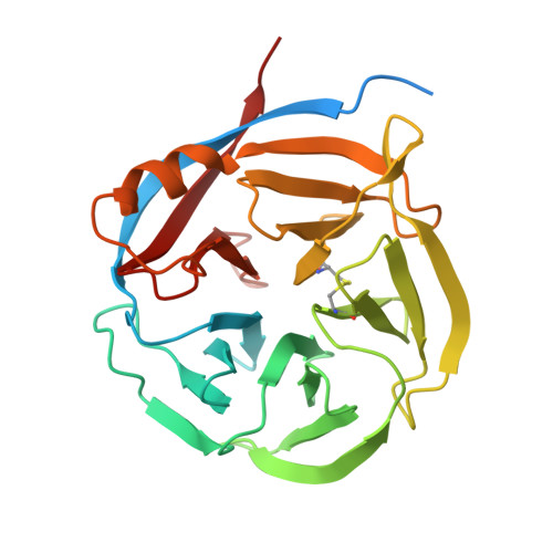
    Although enzymes responsible for the cyclization of amino-terminal glutamine residues are present in both plant and mammal species, none have yet been characterized in bacteria. Based on low sequence homologies to plant glutaminyl cyclases (QCs), we cloned the coding sequences of putative microbial QCs from Zymomonas mobilis (ZmQC) and Myxococcus xanthus (MxQC). The two recombinant enzymes exhibited distinct QC activity, with specificity constants k(cat)/K(m) of 1.47±0.33 mm⁻¹ s⁻¹ (ZmQC) and 142±32.7 mm⁻¹ s⁻¹ (MxQC) towards the fluorescent substrate glutamine-7-amino-4-methyl-coumarine. The measured pH-rate profile of the second order rate constant displayed an interesting deviation towards the acidic limb of the pH chart in the case of ZmQC, whereas MxQC showed maximum activity in the mild alkaline pH range. Analysis of the enzyme variants ZmQCGlu⁴⁶Gln and MxQCGln⁴⁶Glu show that the exchanged residues play a significant role in the pH behaviour of the respective enzymes. In addition, we determined the three dimensional crystal structures of both enzymes. The tertiary structure is defined by a five-bladed β-propeller anchored by a core cation. The structures corroborate the putative location of the active site and confirm the proposed relation between bacterial and plant glutaminyl cyclases.


  • Organizational Affiliation

    Probiodrug AG, Halle/Saale, Germany.


Macromolecules
Find similar proteins by:  (by identity cutoff)  |  3D Structure
Entity ID: 1
MoleculeChains Sequence LengthOrganismDetailsImage
Glutaminyl Cyclase
A, B
268Myxococcus xanthusMutation(s): 0 
EC: 2.3.2.5
UniProt
Find proteins for E7FH78 (Myxococcus xanthus)
Explore E7FH78 
Go to UniProtKB:  E7FH78
Entity Groups  
Sequence Clusters30% Identity50% Identity70% Identity90% Identity95% Identity100% Identity
UniProt GroupE7FH78
Sequence Annotations
Expand
  • Reference Sequence
Small Molecules
Ligands 4 Unique
IDChains Name / Formula / InChI Key2D Diagram3D Interactions
DDQ
Query on DDQ

Download Ideal Coordinates CCD File 
E [auth A],
I [auth B]
DECYLAMINE-N,N-DIMETHYL-N-OXIDE
C12 H27 N O
ZRKZFNZPJKEWPC-UHFFFAOYSA-N
MES
Query on MES

Download Ideal Coordinates CCD File 
D [auth A],
G [auth B]
2-(N-MORPHOLINO)-ETHANESULFONIC ACID
C6 H13 N O4 S
SXGZJKUKBWWHRA-UHFFFAOYSA-N
GOL
Query on GOL

Download Ideal Coordinates CCD File 
H [auth B]GLYCEROL
C3 H8 O3
PEDCQBHIVMGVHV-UHFFFAOYSA-N
CA
Query on CA

Download Ideal Coordinates CCD File 
C [auth A],
F [auth B]
CALCIUM ION
Ca
BHPQYMZQTOCNFJ-UHFFFAOYSA-N
Binding Affinity Annotations 
IDSourceBinding Affinity
DDQ PDBBind:  3NOK IC50: 6.40e+4 (nM) from 1 assay(s)
Experimental Data & Validation

Experimental Data

  • Method: X-RAY DIFFRACTION
  • Resolution: 1.65 Å
  • R-Value Free: 0.236 
  • R-Value Work: 0.195 
  • R-Value Observed: 0.197 
  • Space Group: P 1 21 1
Unit Cell:
Length ( Å )Angle ( ˚ )
a = 65.79α = 90
b = 46.06β = 112.97
c = 81γ = 90
Software Package:
Software NamePurpose
XSCALEdata scaling
PHASERphasing
REFMACrefinement
PDB_EXTRACTdata extraction
MAR345dtbdata collection
XDSdata reduction

Structure Validation

View Full Validation Report


Ligand Structure Quality Assessment 


Entry History 

Deposition Data

Revision History  (Full details and data files)

  • Version 1.0: 2010-11-03
    Type: Initial release
  • Version 1.1: 2011-07-13
    Changes: Version format compliance
  • Version 1.2: 2014-02-26
    Changes: Database references
  • Version 1.3: 2017-11-08
    Changes: Refinement description
  • Version 1.4: 2023-11-01
    Changes: Data collection, Database references, Derived calculations, Refinement description