


The crystal structure of adenine deaminase (AAur1117) from Arthrobacter aurescens
Zhang, Z., Goble, A.M., Raushel, F.M., Swaminathan, S.To be published.
Experimental Data Snapshot
Starting Model: experimental
View more details
wwPDB Validation 3D Report Full Report

Entity ID: 1 | |||||
|---|---|---|---|---|---|
| Molecule | Chains | Sequence Length | Organism | Details | Image |
| Adenosine deaminase 1 | 343 | Paenarthrobacter aurescens TC1 | Mutation(s): 0 Gene Names: add, add1, AAur_1117 EC: 3.5.4.4 (PDB Primary Data), 3.5.4.2 (UniProt) | 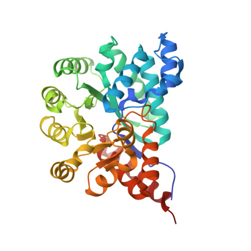 | |
UniProt | |||||
Find proteins for A1R3U3 (Paenarthrobacter aurescens (strain TC1)) Explore A1R3U3 Go to UniProtKB: A1R3U3 | |||||
Entity Groups | |||||
| Sequence Clusters | 30% Identity50% Identity70% Identity90% Identity95% Identity100% Identity | ||||
| UniProt Group | A1R3U3 | ||||
Sequence AnnotationsExpand | |||||
| |||||
| Ligands 2 Unique | |||||
|---|---|---|---|---|---|
| ID | Chains | Name / Formula / InChI Key | 2D Diagram | 3D Interactions | |
| ADE Query on ADE | D [auth A], F [auth B] | ADENINE C5 H5 N5 GFFGJBXGBJISGV-UHFFFAOYSA-N |  | ||
| ZN Query on ZN | C [auth A], E [auth B] | ZINC ION Zn PTFCDOFLOPIGGS-UHFFFAOYSA-N |  | ||
| Length ( Å ) | Angle ( ˚ ) |
|---|---|
| a = 124.104 | α = 90 |
| b = 124.104 | β = 90 |
| c = 89.724 | γ = 90 |
| Software Name | Purpose |
|---|---|
| CBASS | data collection |
| PHASER | phasing |
| PHENIX | refinement |
| HKL-2000 | data reduction |
| HKL-2000 | data scaling |


