4D8D

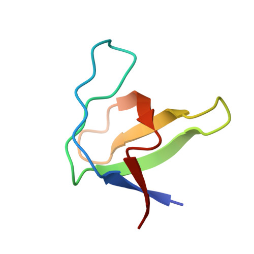
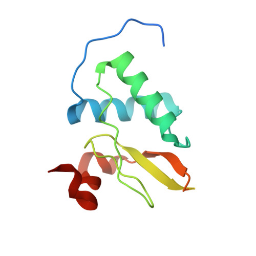
Crystal structure of HIV-1 NEF Fyn-SH3 R96W variant


Experimental Data Snapshot

  • Method: X-RAY DIFFRACTION
  • Resolution: 2.52 Å
  • R-Value Free: 0.238 
  • R-Value Work: 0.212 
  • R-Value Observed: 0.213 

Starting Model: experimental
View more details

wwPDB Validation   3D Report Full Report


This is version 1.3 of the entry. See complete history


Literature

Synergy and allostery in ligand binding by HIV-1 Nef.

Aldehaiman, A.Momin, A.A.Restouin, A.Wang, L.Shi, X.Aljedani, S.Opi, S.Lugari, A.Shahul Hameed, U.F.Ponchon, L.Morelli, X.Huang, M.Dumas, C.Collette, Y.Arold, S.T.

(2021) Biochem J 478: 1525-1545

  • DOI: https://doi.org/10.1042/BCJ20201002
  • Primary Citation of Related Structures:  
    3H0F, 3H0H, 3H0I, 4D8D, 6IPY, 6IPZ, 7D7S

  • PubMed Abstract: 

    The Nef protein of human and simian immunodeficiency viruses boosts viral pathogenicity through its interactions with host cell proteins. By combining the polyvalency of its large unstructured regions with the binding selectivity and strength of its folded core domain, Nef can associate with many different host cell proteins, thereby disrupting their functions. For example, the combination of a linear proline-rich motif and hydrophobic core domain surface allows Nef to bind tightly and specifically to SH3 domains of Src family kinases. We investigated whether the interplay between Nef's flexible regions and its core domain could allosterically influence ligand selection. We found that the flexible regions can associate with the core domain in different ways, producing distinct conformational states that alter the way in which Nef selects for SH3 domains and exposes some of its binding motifs. The ensuing crosstalk between ligands might promote functionally coherent Nef-bound protein ensembles by synergizing certain subsets of ligands while excluding others. We also combined proteomic and bioinformatics analyses to identify human proteins that select SH3 domains in the same way as Nef. We found that only 3% of clones from a whole-human fetal library displayed Nef-like SH3 selectivity. However, in most cases, this selectivity appears to be achieved by a canonical linear interaction rather than by a Nef-like 'tertiary' interaction. Our analysis supports the contention that Nef's mode of hijacking SH3 domains is a virus-specific adaptation with no or very few cellular counterparts. Thus, the Nef tertiary binding surface is a promising virus-specific drug target.


  • Organizational Affiliation

    King Abdullah University of Science and Technology (KAUST), Computational Bioscience Research Center (CBRC), Biological and Environmental Science and Engineering (BESE), Thuwal 23955-6900, Saudi Arabia.


Macromolecules
Find similar proteins by:  (by identity cutoff)  |  3D Structure
Entity ID: 1
MoleculeChains Sequence LengthOrganismDetailsImage
Tyrosine-protein kinase Fyn
A, C
58Homo sapiensMutation(s): 1 
Gene Names: FYNFYN TYROSINE KINASE
EC: 2.7.10.2
UniProt & NIH Common Fund Data Resources
Find proteins for P06241 (Homo sapiens)
Explore P06241 
Go to UniProtKB:  P06241
PHAROS:  P06241
GTEx:  ENSG00000010810 
Entity Groups  
Sequence Clusters30% Identity50% Identity70% Identity90% Identity95% Identity100% Identity
UniProt GroupP06241
Sequence Annotations
Expand
  • Reference Sequence
Find similar proteins by:  (by identity cutoff)  |  3D Structure
Entity ID: 2
MoleculeChains Sequence LengthOrganismDetailsImage
Protein Nef
B, D
151Human immunodeficiency virus type 1 (BRU ISOLATE)Mutation(s): 0 
Gene Names: nef
UniProt
Find proteins for P03406 (Human immunodeficiency virus type 1 group M subtype B (isolate BRU/LAI))
Explore P03406 
Go to UniProtKB:  P03406
Entity Groups  
Sequence Clusters30% Identity50% Identity70% Identity90% Identity95% Identity100% Identity
UniProt GroupP03406
Sequence Annotations
Expand
  • Reference Sequence
Small Molecules
Ligands 1 Unique
IDChains Name / Formula / InChI Key2D Diagram3D Interactions
GOL
Query on GOL

Download Ideal Coordinates CCD File 
E [auth C]GLYCEROL
C3 H8 O3
PEDCQBHIVMGVHV-UHFFFAOYSA-N
Experimental Data & Validation

Experimental Data

  • Method: X-RAY DIFFRACTION
  • Resolution: 2.52 Å
  • R-Value Free: 0.238 
  • R-Value Work: 0.212 
  • R-Value Observed: 0.213 
  • Space Group: P 65 2 2
Unit Cell:
Length ( Å )Angle ( ˚ )
a = 108.01α = 90
b = 108.01β = 90
c = 224.961γ = 120
Software Package:
Software NamePurpose
PHENIXrefinement
PDB_EXTRACTdata extraction
MOSFLMdata reduction
SCALAdata scaling
MOLREPphasing

Structure Validation

View Full Validation Report


Entry History 

Deposition Data

Revision History  (Full details and data files)

  • Version 1.0: 2013-01-16
    Type: Initial release
  • Version 1.1: 2018-01-24
    Changes: Database references
  • Version 1.2: 2021-10-20
    Changes: Database references, Derived calculations
  • Version 1.3: 2023-09-13
    Changes: Data collection, Refinement description