4NGT | pdb_00004ngt

Crystal Structure of Glutamate Carboxypeptidase II in a complex with urea-based inhibitor


Experimental Data Snapshot

  • Method: X-RAY DIFFRACTION
  • Resolution: 2.31 Å
  • R-Value Free: 
    0.223 (Depositor), 0.221 (DCC) 
  • R-Value Work: 
    0.176 (Depositor), 0.176 (DCC) 
  • R-Value Observed: 
    0.178 (Depositor) 

wwPDB Validation   3D Report Full Report


Ligand Structure Quality Assessment 


This is version 2.1 of the entry. See complete history


Literature

Rational design of urea-based glutamate carboxypeptidase II (GCPII) inhibitors as versatile tools for specific drug targeting and delivery.

Tykvart, J.Schimer, J.Barinkova, J.Pachl, P.Postova-Slavetinska, L.Majer, P.Konvalinka, J.Sacha, P.

(2014) Bioorg Med Chem 22: 4099-4108

  • DOI: https://doi.org/10.1016/j.bmc.2014.05.061
  • Primary Citation of Related Structures:  
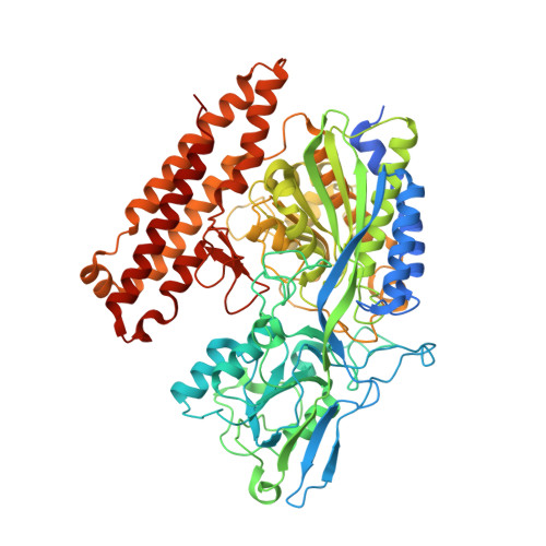
    4NGM, 4NGN, 4NGP, 4NGQ, 4NGR, 4NGS, 4NGT

  • PubMed Abstract: 

    Glutamate carboxypeptidase II (GCPII), also known as prostate specific membrane antigen (PSMA), is an established prostate cancer marker and is considered a promising target for specific anticancer drug delivery. Low-molecular-weight inhibitors of GCPII are advantageous specific ligands for this purpose. However, they must be modified with a linker to enable connection of the ligand with an imaging molecule, anticancer drug, and/or nanocarrier. Here, we describe a structure-activity relationship (SAR) study of GCPII inhibitors with linkers suitable for imaging and drug delivery. Structure-assisted inhibitor design and targeting of a specific GCPII exosite resulted in a 7-fold improvement in Ki value compared to the parent structure. X-ray structural analysis of the inhibitor series led to the identification of several inhibitor binding modes. We also optimized the length of the inhibitor linker for effective attachment to a biotin-binding molecule and showed that the optimized inhibitor could be used to target nanoparticles to cells expressing GCPII.


  • Organizational Affiliation
    • Gilead Sciences and IOCB Research Centre, Institute of Organic Chemistry and Biochemistry, Academy of Sciences of the Czech Republic, v.v.i., Flemingovo n. 2, Prague 6, 166 10 Czech Republic; Department of Biochemistry, Faculty of Natural Science, Charles University, Albertov 6, Prague 2, Czech Republic.

Macromolecules
Find similar proteins by:  (by identity cutoff)  |  3D Structure
Entity ID: 1
MoleculeChains Sequence LengthOrganismDetailsImage
Glutamate carboxypeptidase 2739Homo sapiensMutation(s): 0 
Gene Names: FOLHFOLH1GIG27NAALAD1PSMPSMA
EC: 3.4.17.21
UniProt & NIH Common Fund Data Resources
Find proteins for Q04609 (Homo sapiens)
Explore Q04609 
Go to UniProtKB:  Q04609
PHAROS:  Q04609
GTEx:  ENSG00000086205 
Entity Groups  
Sequence Clusters30% Identity50% Identity70% Identity90% Identity95% Identity100% Identity
UniProt GroupQ04609
Glycosylation
Glycosylation Sites: 6Go to GlyGen: Q04609-1
Sequence Annotations
Expand
  • Reference Sequence
Oligosaccharides

Help

Entity ID: 2
MoleculeChains Length2D Diagram Glycosylation3D Interactions
2-acetamido-2-deoxy-beta-D-glucopyranose-(1-4)-2-acetamido-2-deoxy-beta-D-glucopyranose
B
2N-Glycosylation
Glycosylation Resources
GlyTouCan:  G42666HT
GlyCosmos:  G42666HT
GlyGen:  G42666HT
Entity ID: 3
MoleculeChains Length2D Diagram Glycosylation3D Interactions
beta-D-mannopyranose-(1-4)-2-acetamido-2-deoxy-beta-D-glucopyranose-(1-4)-2-acetamido-2-deoxy-beta-D-glucopyranose
C
3N-Glycosylation
Glycosylation Resources
GlyTouCan:  G15407YE
GlyCosmos:  G15407YE
GlyGen:  G15407YE
Small Molecules
Ligands 6 Unique
IDChains Name / Formula / InChI Key2D Diagram3D Interactions
J42
Query on J42

Download Ideal Coordinates CCD File 
M [auth A]N-{[(1S)-5-({[4-bromo-2-({4,44-dioxo-48-[(3aS,4S,6aR)-2-oxohexahydro-1H-thieno[3,4-d]imidazol-4-yl]-7,10,13,16,19,22,25,28,31,34,37,40-dodecaoxa-3,43-diazaoctatetracont-1-yl}oxy)phenyl]carbamoyl}amino)-1-carboxypentyl]carbamoyl}-L-glutamic acid
C58 H97 Br N8 O24 S
GEHJWUZNJRAVQB-HJXABITDSA-N
NAG
Query on NAG

Download Ideal Coordinates CCD File 
D [auth A],
E [auth A],
F [auth A],
G [auth A]
2-acetamido-2-deoxy-beta-D-glucopyranose
C8 H15 N O6
OVRNDRQMDRJTHS-FMDGEEDCSA-N
ZN
Query on ZN

Download Ideal Coordinates CCD File 
H [auth A],
I [auth A]
ZINC ION
Zn
PTFCDOFLOPIGGS-UHFFFAOYSA-N
CA
Query on CA

Download Ideal Coordinates CCD File 
K [auth A]CALCIUM ION
Ca
BHPQYMZQTOCNFJ-UHFFFAOYSA-N
CL
Query on CL

Download Ideal Coordinates CCD File 
J [auth A]CHLORIDE ION
Cl
VEXZGXHMUGYJMC-UHFFFAOYSA-M
NA
Query on NA

Download Ideal Coordinates CCD File 
L [auth A]SODIUM ION
Na
FKNQFGJONOIPTF-UHFFFAOYSA-N
Binding Affinity Annotations 
IDSourceBinding Affinity
J42 BindingDB:  4NGT Ki: min: 0.22, max: 3.9 (nM) from 2 assay(s)
Experimental Data & Validation

Experimental Data

  • Method: X-RAY DIFFRACTION
  • Resolution: 2.31 Å
  • R-Value Free:  0.223 (Depositor), 0.221 (DCC) 
  • R-Value Work:  0.176 (Depositor), 0.176 (DCC) 
  • R-Value Observed: 0.178 (Depositor) 
Space Group: I 2 2 2
Unit Cell:
Length ( Å )Angle ( ˚ )
a = 101.543α = 90
b = 130.297β = 90
c = 159.864γ = 90
Software Package:
Software NamePurpose
XSCALEdata scaling
MOLREPphasing
REFMACrefinement
PDB_EXTRACTdata extraction
XDSdata reduction
XDSdata scaling

Structure Validation

View Full Validation Report


Ligand Structure Quality Assessment 


Entry History 

Deposition Data

Revision History  (Full details and data files)

  • Version 1.0: 2014-06-18
    Type: Initial release
  • Version 1.1: 2019-12-18
    Changes: Database references, Derived calculations
  • Version 2.0: 2020-07-29
    Type: Remediation
    Reason: Carbohydrate remediation
    Changes: Advisory, Atomic model, Data collection, Derived calculations, Structure summary
  • Version 2.1: 2024-11-20
    Changes: Data collection, Database references, Structure summary