4O8H

0.85A resolution structure of PEG 400 Bound Cyclophilin D


Experimental Data Snapshot

  • Method: X-RAY DIFFRACTION
  • Resolution: 0.85 Å
  • R-Value Free: 0.119 
  • R-Value Work: 0.107 
  • R-Value Observed: 0.108 

wwPDB Validation   3D Report Full Report


This is version 1.3 of the entry. See complete history


Literature

High-resolution crystal structures of two crystal forms of human cyclophilin D in complex with PEG 400 molecules.

Valasani, K.R.Carlson, E.A.Battaile, K.P.Bisson, A.Wang, C.Lovell, S.ShiDu Yan, S.

(2014) Acta Crystallogr Sect F Struct Biol Cryst Commun 70: 717-722

  • DOI: https://doi.org/10.1107/S2053230X14009480
  • Primary Citation of Related Structures:  
    4O8H, 4O8I

  • PubMed Abstract: 

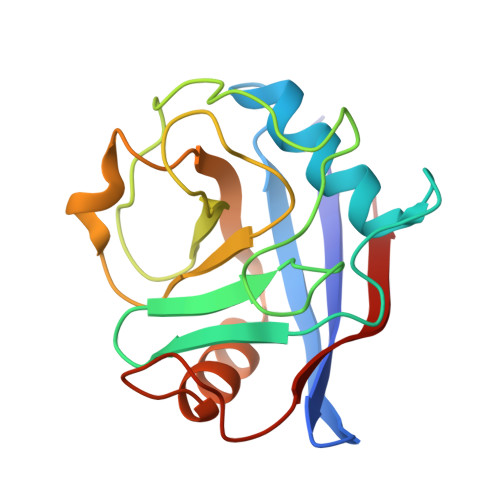
    Cyclophilin D (CypD) is a key mitochondrial target for amyloid-β-induced mitochondrial and synaptic dysfunction and is considered a potential drug target for Alzheimer's disease. The high-resolution crystal structures of primitive orthorhombic (CypD-o) and primitive tetragonal (CypD-t) forms have been determined to 1.45 and 0.85 Å resolution, respectively, and are nearly identical structurally. Although an isomorphous structure of CypD-t has previously been reported, the structure reported here was determined at atomic resolution, while CypD-o represents a new crystal form for this protein. In addition, each crystal form contains a PEG 400 molecule bound to the same region along with a second PEG 400 site in CypD-t which occupies the cyclosporine A inhibitor binding site of CypD. Highly precise structural information for CypD should be extremely useful for discerning the detailed interaction of small molecules, particularly drugs and/or inhibitors, bound to CypD. The 0.85 Å resolution structure of CypD-t is the highest to date for any CypD structure.


  • Organizational Affiliation

    Department of Pharmacology and Toxicology and Higuchi Bioscience Center, University of Kansas, Lawrence, KS 66047, USA.


Macromolecules
Find similar proteins by:  (by identity cutoff)  |  3D Structure
Entity ID: 1
MoleculeChains Sequence LengthOrganismDetailsImage
Peptidyl-prolyl cis-trans isomerase F, mitochondrial165Homo sapiensMutation(s): 1 
Gene Names: PPIFCYP3
EC: 5.2.1.8
UniProt & NIH Common Fund Data Resources
Find proteins for P30405 (Homo sapiens)
Explore P30405 
Go to UniProtKB:  P30405
PHAROS:  P30405
GTEx:  ENSG00000108179 
Entity Groups  
Sequence Clusters30% Identity50% Identity70% Identity90% Identity95% Identity100% Identity
UniProt GroupP30405
Sequence Annotations
Expand
  • Reference Sequence
Small Molecules
Ligands 2 Unique
IDChains Name / Formula / InChI Key2D Diagram3D Interactions
1PE
Query on 1PE

Download Ideal Coordinates CCD File 
C [auth A]PENTAETHYLENE GLYCOL
C10 H22 O6
JLFNLZLINWHATN-UHFFFAOYSA-N
PEG
Query on PEG

Download Ideal Coordinates CCD File 
B [auth A]DI(HYDROXYETHYL)ETHER
C4 H10 O3
MTHSVFCYNBDYFN-UHFFFAOYSA-N
Experimental Data & Validation

Experimental Data

  • Method: X-RAY DIFFRACTION
  • Resolution: 0.85 Å
  • R-Value Free: 0.119 
  • R-Value Work: 0.107 
  • R-Value Observed: 0.108 
  • Space Group: P 41 21 2
Unit Cell:
Length ( Å )Angle ( ˚ )
a = 57.02α = 90
b = 57.02β = 90
c = 87.159γ = 90
Software Package:
Software NamePurpose
Aimlessdata scaling
PHASERphasing
PHENIXrefinement
PDB_EXTRACTdata extraction
JDirectordata collection
XSCALEdata scaling

Structure Validation

View Full Validation Report


Entry History 

Deposition Data

Revision History  (Full details and data files)

  • Version 1.0: 2014-06-11
    Type: Initial release
  • Version 1.1: 2014-09-24
    Changes: Database references
  • Version 1.2: 2017-11-22
    Changes: Refinement description
  • Version 1.3: 2023-09-20
    Changes: Data collection, Database references, Derived calculations, Refinement description