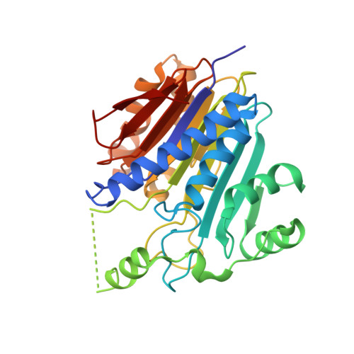
4OSX

STRUCTURE of UNCLEAVED GLYCINE-BOUND HUMAN L-ASPARAGINASE PROTEIN


Experimental Data Snapshot

  • Method: X-RAY DIFFRACTION
  • Resolution: 1.95 Å
  • R-Value Free: 0.209 
  • R-Value Work: 0.167 
  • R-Value Observed: 0.169 

wwPDB Validation   3D Report Full Report


This is version 1.1 of the entry. See complete history


Literature

Free Glycine Accelerates the Autoproteolytic Activation of Human Asparaginase.

Su, Y.Karamitros, C.S.Nomme, J.Mcsorley, T.Konrad, M.Lavie, A.

(2013) Chem Biol 20: 533

  • DOI: https://doi.org/10.1016/j.chembiol.2013.03.006
  • Primary Citation of Related Structures:  
    4OSX, 4OSY

  • PubMed Abstract: 

    Human asparaginase 3 (hASNase3), which belongs to the N-terminal nucleophile hydrolase superfamily, is synthesized as a single polypeptide that is devoid of asparaginase activity. Intramolecular autoproteolytic processing releases the amino group of Thr168, a moiety required for catalyzing asparagine hydrolysis. Recombinant hASNase3 purifies as the uncleaved, asparaginase-inactive form and undergoes self-cleavage to the active form at a very slow rate. Here, we show that the free amino acid glycine selectively acts to accelerate hASNase3 cleavage both in vitro and in human cells. Other small amino acids such as alanine, serine, or the substrate asparagine are not capable of promoting autoproteolysis. Crystal structures of hASNase3 in complex with glycine in the uncleaved and cleaved enzyme states reveal the mechanism of glycine-accelerated posttranslational processing and explain why no other amino acid can substitute for glycine.


  • Organizational Affiliation

    Department of Biochemistry and Molecular Genetics, University of Illinois at Chicago, Chicago, IL 60607, USA.


Macromolecules
Find similar proteins by:  (by identity cutoff)  |  3D Structure
Entity ID: 1
MoleculeChains Sequence LengthOrganismDetailsImage
Isoaspartyl peptidase/L-asparaginase
A, B
309Homo sapiensMutation(s): 0 
Gene Names: ALPASRGL1CRASH
EC: 3.4.19.5 (PDB Primary Data), 3.5.1.1 (PDB Primary Data)
UniProt & NIH Common Fund Data Resources
Find proteins for Q7L266 (Homo sapiens)
Explore Q7L266 
Go to UniProtKB:  Q7L266
PHAROS:  Q7L266
GTEx:  ENSG00000162174 
Entity Groups  
Sequence Clusters30% Identity50% Identity70% Identity90% Identity95% Identity100% Identity
UniProt GroupQ7L266
Sequence Annotations
Expand
  • Reference Sequence
Experimental Data & Validation

Experimental Data

  • Method: X-RAY DIFFRACTION
  • Resolution: 1.95 Å
  • R-Value Free: 0.209 
  • R-Value Work: 0.167 
  • R-Value Observed: 0.169 
  • Space Group: P 65
Unit Cell:
Length ( Å )Angle ( ˚ )
a = 59.94α = 90
b = 59.94β = 90
c = 301.1γ = 120
Software Package:
Software NamePurpose
CrystalCleardata collection
MOLREPphasing
REFMACrefinement
XDSdata reduction
XDSdata scaling

Structure Validation

View Full Validation Report


Entry History 

Deposition Data

Revision History  (Full details and data files)

  • Version 1.0: 2014-03-05
    Type: Initial release
  • Version 1.1: 2023-09-20
    Changes: Data collection, Database references, Derived calculations, Refinement description