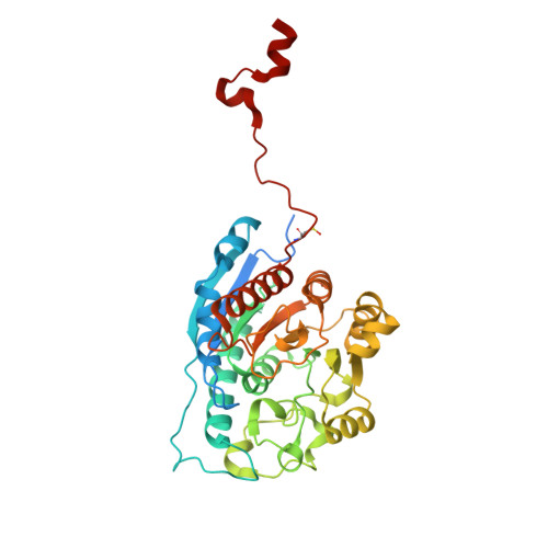
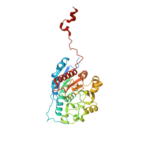
4Q3R

Crystal structure of Schistosoma mansoni arginase in complex with inhibitor ABHDP


Experimental Data Snapshot

  • Method: X-RAY DIFFRACTION
  • Resolution: 2.17 Å
  • R-Value Free: 0.206 
  • R-Value Work: 0.174 
  • R-Value Observed: 0.176 

wwPDB Validation   3D Report Full Report


Ligand Structure Quality Assessment 


This is version 1.4 of the entry. See complete history


Literature

Crystal Structure of Schistosoma mansoni Arginase, a Potential Drug Target for the Treatment of Schistosomiasis.

Hai, Y.Edwards, J.E.Van Zandt, M.C.Hoffmann, K.F.Christianson, D.W.

(2014) Biochemistry 53: 4671-4684

  • DOI: https://doi.org/10.1021/bi5004519
  • Primary Citation of Related Structures:  
    4Q3P, 4Q3Q, 4Q3R, 4Q3S, 4Q3T, 4Q3U, 4Q3V, 4Q40, 4Q41, 4Q42

  • PubMed Abstract: 

    The X-ray crystal structure of arginase from Schistosoma mansoni (SmARG) and the structures of its complexes with several amino acid inhibitors have been determined at atomic resolution. SmARG is a binuclear manganese metalloenzyme that catalyzes the hydrolysis of l-arginine to form l-ornithine and urea, and this enzyme is upregulated in all forms of the parasite that interact with the human host. Current hypotheses suggest that parasitic arginases could play a role in host immune evasion by depleting pools of substrate l-arginine that would otherwise be utilized for NO biosynthesis and NO-dependent processes in the immune response. Although the amino acid sequence of SmARG is only 42% identical with that of human arginase I, residues important for substrate binding and catalysis are strictly conserved. In general, classical amino acid inhibitors such as 2(S)-amino-6-boronohexanoic acid (ABH) tend to bind more weakly to SmARG than to human arginase I despite identical inhibitor binding modes in each enzyme active site. The identification of a patch on the enzyme surface capable of accommodating the additional Cα substitutent of an α,α-disubstituted amino acid inhibitor suggests that such inhibitors could exhibit higher affinity and biological activity. The structures of SmARG complexed with two different α,α-disubstituted derivatives of ABH are presented and provide a proof of concept for this approach in the enhancement of enzyme-inhibitor affinity.


  • Organizational Affiliation

    Roy and Diana Vagelos Laboratories, Department of Chemistry, University of Pennsylvania , Philadelphia, Pennsylvania 19104-6323, United States.


Macromolecules
Find similar proteins by:  (by identity cutoff)  |  3D Structure
Entity ID: 1
MoleculeChains Sequence LengthOrganismDetailsImage
Arginase
A, B
385Schistosoma mansoniMutation(s): 0 
Gene Names: Smp_059980
EC: 3.5.3.1
UniProt
Find proteins for Q6WVP6 (Schistosoma mansoni)
Explore Q6WVP6 
Go to UniProtKB:  Q6WVP6
Entity Groups  
Sequence Clusters30% Identity50% Identity70% Identity90% Identity95% Identity100% Identity
UniProt GroupQ6WVP6
Sequence Annotations
Expand
  • Reference Sequence
Find similar proteins by:  (by identity cutoff)  |  3D Structure
Entity ID: 2
MoleculeChains Sequence LengthOrganismDetailsImage
Arginase
C, D
385Schistosoma mansoniMutation(s): 0 
Gene Names: Smp_059980
EC: 3.5.3.1
UniProt
Find proteins for Q6WVP6 (Schistosoma mansoni)
Explore Q6WVP6 
Go to UniProtKB:  Q6WVP6
Entity Groups  
Sequence Clusters30% Identity50% Identity70% Identity90% Identity95% Identity100% Identity
UniProt GroupQ6WVP6
Sequence Annotations
Expand
  • Reference Sequence
Small Molecules
Ligands 6 Unique
IDChains Name / Formula / InChI Key2D Diagram3D Interactions
XA2
Query on XA2

Download Ideal Coordinates CCD File 
FA [auth C],
K [auth A],
LA [auth D],
W [auth B]
(R)-2-amino-6-borono-2-(1-(3,4-dichlorobenzyl)piperidin-4-yl)hexanoic acid
C18 H28 B Cl2 N2 O5
LYUGMNLZENMUTE-GOSISDBHSA-N
PGE
Query on PGE

Download Ideal Coordinates CCD File 
N [auth A]TRIETHYLENE GLYCOL
C6 H14 O4
ZIBGPFATKBEMQZ-UHFFFAOYSA-N
CS
Query on CS

Download Ideal Coordinates CCD File 
BA [auth C]
CA [auth C]
EA [auth C]
G [auth A]
H [auth A]
BA [auth C],
CA [auth C],
EA [auth C],
G [auth A],
H [auth A],
I [auth A],
KA [auth D],
Q [auth B],
R [auth B],
S [auth B],
T [auth B]
CESIUM ION
Cs
NCMHKCKGHRPLCM-UHFFFAOYSA-N
GOL
Query on GOL

Download Ideal Coordinates CCD File 
GA [auth C]
HA [auth C]
L [auth A]
M [auth A]
X [auth B]
GA [auth C],
HA [auth C],
L [auth A],
M [auth A],
X [auth B],
Y [auth B]
GLYCEROL
C3 H8 O3
PEDCQBHIVMGVHV-UHFFFAOYSA-N
MN
Query on MN

Download Ideal Coordinates CCD File 
AA [auth C]
E [auth A]
F [auth A]
IA [auth D]
JA [auth D]
AA [auth C],
E [auth A],
F [auth A],
IA [auth D],
JA [auth D],
O [auth B],
P [auth B],
Z [auth C]
MANGANESE (II) ION
Mn
WAEMQWOKJMHJLA-UHFFFAOYSA-N
CL
Query on CL

Download Ideal Coordinates CCD File 
DA [auth C],
J [auth A],
U [auth B],
V [auth B]
CHLORIDE ION
Cl
VEXZGXHMUGYJMC-UHFFFAOYSA-M
Modified Residues  1 Unique
IDChains TypeFormula2D DiagramParent
CSO
Query on CSO
A, B
L-PEPTIDE LINKINGC3 H7 N O3 SCYS
Binding Affinity Annotations 
IDSourceBinding Affinity
XA2 Binding MOAD:  4Q3R Kd: 540 (nM) from 1 assay(s)
Experimental Data & Validation

Experimental Data

  • Method: X-RAY DIFFRACTION
  • Resolution: 2.17 Å
  • R-Value Free: 0.206 
  • R-Value Work: 0.174 
  • R-Value Observed: 0.176 
  • Space Group: P 21 3
Unit Cell:
Length ( Å )Angle ( ˚ )
a = 177.488α = 90
b = 177.488β = 90
c = 177.488γ = 90
Software Package:
Software NamePurpose
CBASSdata collection
PHASERphasing
PHENIXrefinement
HKL-2000data reduction
HKL-2000data scaling

Structure Validation

View Full Validation Report


Ligand Structure Quality Assessment 


Entry History 

Deposition Data

Revision History  (Full details and data files)

  • Version 1.0: 2014-07-16
    Type: Initial release
  • Version 1.1: 2014-07-23
    Changes: Database references
  • Version 1.2: 2014-08-06
    Changes: Database references
  • Version 1.3: 2023-09-20
    Changes: Data collection, Database references, Derived calculations, Refinement description
  • Version 1.4: 2023-12-06
    Changes: Data collection