4CK7

Pseudo-atomic model of microtubule-bound human kinesin-5 motor domain in presence of adp.alfx (NECK-LINKER IN ITS DISCONNECTED CONFORMATION, based on cryo-electron microscopy experiment


Experimental Data Snapshot

  • Method: ELECTRON MICROSCOPY
  • Resolution: 9.20 Å
  • Aggregation State: PARTICLE 
  • Reconstruction Method: SINGLE PARTICLE 

wwPDB Validation   3D Report Full Report


This is version 1.2 of the entry. See complete history


Literature

Comprehensive Structural Model of the Mechanochemical Cycle of a Mitotic Motor Highlights Molecular Adaptations in the Kinesin Family.

Goulet, A.Major, J.Jun, Y.Gross, S.P.Rosenfeld, S.S.Moores, C.A.

(2014) Proc Natl Acad Sci U S A 111: 1837

  • DOI: https://doi.org/10.1073/pnas.1319848111
  • Primary Citation of Related Structures:  
    4CK5, 4CK6, 4CK7

  • PubMed Abstract: 

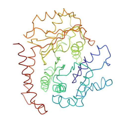
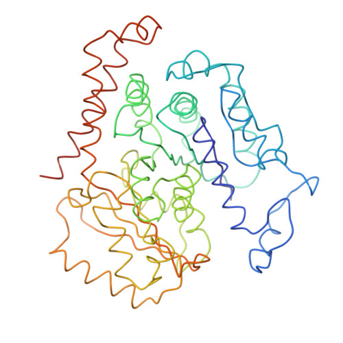
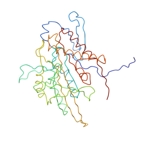
    Kinesins are responsible for a wide variety of microtubule-based, ATP-dependent functions. Their motor domain drives these activities, but the molecular adaptations that specify these diverse and essential cellular activities are poorly understood. It has been assumed that the first identified kinesin--the transport motor kinesin-1--is the mechanistic paradigm for the entire superfamily, but accumulating evidence suggests otherwise. To address the deficits in our understanding of the molecular basis of functional divergence within the kinesin superfamily, we studied kinesin-5s, which are essential mitotic motors whose inhibition blocks cell division. Using cryo-electron microscopy and determination of structure at subnanometer resolution, we have visualized conformations of microtubule-bound human kinesin-5 motor domain at successive steps in its ATPase cycle. After ATP hydrolysis, nucleotide-dependent conformational changes in the active site are allosterically propagated into rotations of the motor domain and uncurling of the drug-binding loop L5. In addition, the mechanical neck-linker element that is crucial for motor stepping undergoes discrete, ordered displacements. We also observed large reorientations of the motor N terminus that indicate its importance for kinesin-5 function through control of neck-linker conformation. A kinesin-5 mutant lacking this N terminus is enzymatically active, and ATP-dependent neck-linker movement and motility are defective, although not ablated. All these aspects of kinesin-5 mechanochemistry are distinct from kinesin-1. Our findings directly demonstrate the regulatory role of the kinesin-5 N terminus in collaboration with the motor's structured neck-linker and highlight the multiple adaptations within kinesin motor domains that tune their mechanochemistries according to distinct functional requirements.


  • Organizational Affiliation

    Institute of Structural and Molecular Biology, Birkbeck College, London, WC1E 7HX, United Kingdon.


Macromolecules
Find similar proteins by:  (by identity cutoff)  |  3D Structure
Entity ID: 1
MoleculeChains Sequence LengthOrganismDetailsImage
TUBULIN ALPHA-1D CHAIN452Bos taurusMutation(s): 0 
UniProt
Find proteins for Q2HJ86 (Bos taurus)
Explore Q2HJ86 
Go to UniProtKB:  Q2HJ86
Entity Groups  
Sequence Clusters30% Identity50% Identity70% Identity90% Identity95% Identity100% Identity
UniProt GroupQ2HJ86
Sequence Annotations
Expand
  • Reference Sequence
Find similar proteins by:  (by identity cutoff)  |  3D Structure
Entity ID: 2
MoleculeChains Sequence LengthOrganismDetailsImage
TUBULIN BETA-2B CHAIN445Bos taurusMutation(s): 0 
UniProt
Find proteins for Q6B856 (Bos taurus)
Explore Q6B856 
Go to UniProtKB:  Q6B856
Entity Groups  
Sequence Clusters30% Identity50% Identity70% Identity90% Identity95% Identity100% Identity
UniProt GroupQ6B856
Sequence Annotations
Expand
  • Reference Sequence
Find similar proteins by:  (by identity cutoff)  |  3D Structure
Entity ID: 3
MoleculeChains Sequence LengthOrganismDetailsImage
KINESIN-LIKE PROTEIN KIF11373Homo sapiensMutation(s): 1 
UniProt & NIH Common Fund Data Resources
Find proteins for P52732 (Homo sapiens)
Explore P52732 
Go to UniProtKB:  P52732
PHAROS:  P52732
GTEx:  ENSG00000138160 
Entity Groups  
Sequence Clusters30% Identity50% Identity70% Identity90% Identity95% Identity100% Identity
UniProt GroupP52732
Sequence Annotations
Expand
  • Reference Sequence
Small Molecules
Ligands 6 Unique
IDChains Name / Formula / InChI Key2D Diagram3D Interactions
TA1
Query on TA1

Download Ideal Coordinates CCD File 
G [auth B]TAXOL
C47 H51 N O14
RCINICONZNJXQF-MZXODVADSA-N
GTP
Query on GTP

Download Ideal Coordinates CCD File 
E [auth A]GUANOSINE-5'-TRIPHOSPHATE
C10 H16 N5 O14 P3
XKMLYUALXHKNFT-UUOKFMHZSA-N
GDP
Query on GDP

Download Ideal Coordinates CCD File 
F [auth B]GUANOSINE-5'-DIPHOSPHATE
C10 H15 N5 O11 P2
QGWNDRXFNXRZMB-UUOKFMHZSA-N
ADP
Query on ADP

Download Ideal Coordinates CCD File 
J [auth C]ADENOSINE-5'-DIPHOSPHATE
C10 H15 N5 O10 P2
XTWYTFMLZFPYCI-KQYNXXCUSA-N
AF3
Query on AF3

Download Ideal Coordinates CCD File 
I [auth C]ALUMINUM FLUORIDE
Al F3
KLZUFWVZNOTSEM-UHFFFAOYSA-K
MG
Query on MG

Download Ideal Coordinates CCD File 
D [auth A],
H [auth C]
MAGNESIUM ION
Mg
JLVVSXFLKOJNIY-UHFFFAOYSA-N
Binding Affinity Annotations 
IDSourceBinding Affinity
TA1 BindingDB:  4CK7 EC50: 520 (nM) from 1 assay(s)
Experimental Data & Validation

Experimental Data

  • Method: ELECTRON MICROSCOPY
  • Resolution: 9.20 Å
  • Aggregation State: PARTICLE 
  • Reconstruction Method: SINGLE PARTICLE 
EM Software:
TaskSoftware PackageVersion
RECONSTRUCTIONFREALIGN
RECONSTRUCTIONSPIDER

Structure Validation

View Full Validation Report


Entry History 

Deposition Data

Revision History  (Full details and data files)

  • Version 1.0: 2014-02-05
    Type: Initial release
  • Version 1.1: 2014-02-19
    Changes: Database references
  • Version 1.2: 2017-08-30
    Changes: Data collection, Refinement description