4NOG

Crystal structure of a putative ornithine aminotransferase from Toxoplasma gondii ME49 in complex with pyrodoxal-5'-phosphate


Experimental Data Snapshot

  • Method: X-RAY DIFFRACTION
  • Resolution: 1.20 Å
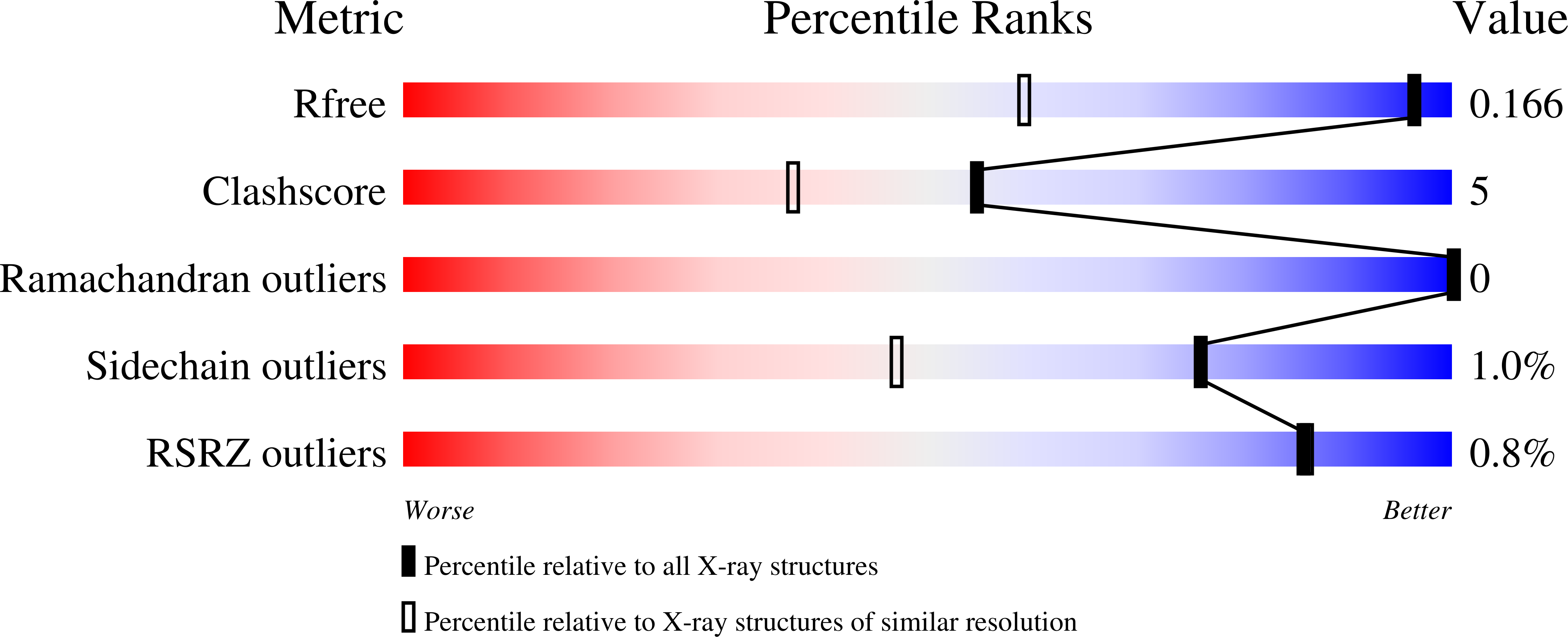
  • R-Value Free: 0.165 
  • R-Value Work: 0.133 
  • R-Value Observed: 0.135 

Starting Model: experimental
View more details

wwPDB Validation   3D Report Full Report


Ligand Structure Quality Assessment 


This is version 1.4 of the entry. See complete history


Literature

CSGID Solves Structures and Identifies Phenotypes for Five Enzymes in Toxoplasma gondii .

Lykins, J.D.Filippova, E.V.Halavaty, A.S.Minasov, G.Zhou, Y.Dubrovska, I.Flores, K.J.Shuvalova, L.A.Ruan, J.El Bissati, K.Dovgin, S.Roberts, C.W.Woods, S.Moulton, J.D.Moulton, H.McPhillie, M.J.Muench, S.P.Fishwick, C.W.G.Sabini, E.Shanmugam, D.Roos, D.S.McLeod, R.Anderson, W.F.Ngo, H.M.

(2018) Front Cell Infect Microbiol 8: 352-352

  • DOI: https://doi.org/10.3389/fcimb.2018.00352
  • Primary Citation of Related Structures:  
    4NML, 4NOG, 4NU7, 4O0N, 4ODI, 5BXI

  • PubMed Abstract: 

    Toxoplasma gondii , an Apicomplexan parasite, causes significant morbidity and mortality, including severe disease in immunocompromised hosts and devastating congenital disease, with no effective treatment for the bradyzoite stage. To address this, we used the Tropical Disease Research database, crystallography, molecular modeling, and antisense to identify and characterize a range of potential therapeutic targets for toxoplasmosis. Phosphoglycerate mutase II (PGMII), nucleoside diphosphate kinase (NDK), ribulose phosphate 3-epimerase (RPE), ribose-5-phosphate isomerase (RPI), and ornithine aminotransferase (OAT) were structurally characterized. Crystallography revealed insights into the overall structure, protein oligomeric states and molecular details of active sites important for ligand recognition. Literature and molecular modeling suggested potential inhibitors and druggability. The targets were further studied with vivoPMO to interrupt enzyme synthesis, identifying the targets as potentially important to parasitic replication and, therefore, of therapeutic interest. Targeted vivoPMO resulted in statistically significant perturbation of parasite replication without concomitant host cell toxicity, consistent with a previous CRISPR/Cas9 screen showing PGM, RPE, and RPI contribute to parasite fitness. PGM, RPE, and RPI have the greatest promise for affecting replication in tachyzoites. These targets are shared between other medically important parasites and may have wider therapeutic potential.


  • Organizational Affiliation

    Pritzker School of Medicine, University of Chicago, Chicago, IL, United States.


Macromolecules
Find similar proteins by:  (by identity cutoff)  |  3D Structure
Entity ID: 1
MoleculeChains Sequence LengthOrganismDetailsImage
Putative ornithine aminotransferase, mitochondrial
A, B
442Toxoplasma gondii ME49Mutation(s): 0 
Gene Names: TGME49_069110TGME49_269110
EC: 2.6.1.13
UniProt
Find proteins for S8EY38 (Toxoplasma gondii (strain ATCC 50611 / Me49))
Explore S8EY38 
Go to UniProtKB:  S8EY38
Entity Groups  
Sequence Clusters30% Identity50% Identity70% Identity90% Identity95% Identity100% Identity
UniProt GroupS8EY38
Sequence Annotations
Expand
  • Reference Sequence
Small Molecules
Ligands 5 Unique
IDChains Name / Formula / InChI Key2D Diagram3D Interactions
PLP
Query on PLP

Download Ideal Coordinates CCD File 
C [auth A],
E [auth B]
PYRIDOXAL-5'-PHOSPHATE
C8 H10 N O6 P
NGVDGCNFYWLIFO-UHFFFAOYSA-N
BTB
Query on BTB

Download Ideal Coordinates CCD File 
G [auth B]2-[BIS-(2-HYDROXY-ETHYL)-AMINO]-2-HYDROXYMETHYL-PROPANE-1,3-DIOL
C8 H19 N O5
OWMVSZAMULFTJU-UHFFFAOYSA-N
PEG
Query on PEG

Download Ideal Coordinates CCD File 
H [auth B]DI(HYDROXYETHYL)ETHER
C4 H10 O3
MTHSVFCYNBDYFN-UHFFFAOYSA-N
BME
Query on BME

Download Ideal Coordinates CCD File 
D [auth A],
I [auth B]
BETA-MERCAPTOETHANOL
C2 H6 O S
DGVVWUTYPXICAM-UHFFFAOYSA-N
ACT
Query on ACT

Download Ideal Coordinates CCD File 
F [auth B]ACETATE ION
C2 H3 O2
QTBSBXVTEAMEQO-UHFFFAOYSA-M
Experimental Data & Validation

Experimental Data

  • Method: X-RAY DIFFRACTION
  • Resolution: 1.20 Å
  • R-Value Free: 0.165 
  • R-Value Work: 0.133 
  • R-Value Observed: 0.135 
  • Space Group: P 1
  • Diffraction Data: https://doi.org/10.18430/m34nog
Unit Cell:
Length ( Å )Angle ( ˚ )
a = 56.19α = 100.58
b = 61.328β = 93.23
c = 63.678γ = 107.74
Software Package:
Software NamePurpose
Blu-Icedata collection
PHASERphasing
REFMACrefinement
HKL-3000data reduction
HKL-3000data scaling

Structure Validation

View Full Validation Report


Ligand Structure Quality Assessment 


Entry History 

Revision History  (Full details and data files)

  • Version 1.0: 2013-12-04
    Type: Initial release
  • Version 1.1: 2017-11-22
    Changes: Refinement description
  • Version 1.2: 2018-01-24
    Changes: Database references, Structure summary
  • Version 1.3: 2021-01-27
    Changes: Database references, Derived calculations
  • Version 1.4: 2023-09-20
    Changes: Data collection, Database references, Refinement description