5A3C

Crystal structure of the ADP-ribosylating sirtuin (SirTM) from Streptococcus pyogenes in complex with NAD


Experimental Data Snapshot

  • Method: X-RAY DIFFRACTION
  • Resolution: 2.03 Å
  • R-Value Free: 0.233 
  • R-Value Work: 0.176 
  • R-Value Observed: 0.178 

wwPDB Validation   3D Report Full Report


Ligand Structure Quality Assessment 


This is version 1.1 of the entry. See complete history


Literature

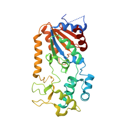
Identification of a Class of Protein Adp-Ribosylating Sirtuins in Microbial Pathogens.

Rack, J.G.Morra, R.Barkauskaite, E.Kraehenbuehl, R.Ariza, A.Qu, Y.Ortmayer, M.Leidecker, O.Cameron, D.R.Matic, I.Peleg, A.Y.Leys, D.Traven, A.Ahel, I.

(2015) Mol Cell 59: 309

  • DOI: https://doi.org/10.1016/j.molcel.2015.06.013
  • Primary Citation of Related Structures:  
    5A35, 5A3A, 5A3B, 5A3C

  • PubMed Abstract: 

    Sirtuins are an ancient family of NAD(+)-dependent deacylases connected with the regulation of fundamental cellular processes including metabolic homeostasis and genome integrity. We show the existence of a hitherto unrecognized class of sirtuins, found predominantly in microbial pathogens. In contrast to earlier described classes, these sirtuins exhibit robust protein ADP-ribosylation activity. In our model organisms, Staphylococcus aureus and Streptococcus pyogenes, the activity is dependent on prior lipoylation of the target protein and can be reversed by a sirtuin-associated macrodomain protein. Together, our data describe a sirtuin-dependent reversible protein ADP-ribosylation system and establish a crosstalk between lipoylation and mono-ADP-ribosylation. We propose that these posttranslational modifications modulate microbial virulence by regulating the response to host-derived reactive oxygen species.


  • Organizational Affiliation

    Sir William Dunn School of Pathology, University of Oxford, South Parks Road, Oxford OX1 3RE, UK.


Macromolecules
Find similar proteins by:  (by identity cutoff)  |  3D Structure
Entity ID: 1
MoleculeChains Sequence LengthOrganismDetailsImage
SIR2 FAMILY PROTEIN303Streptococcus pyogenes str. ManfredoMutation(s): 0 
EC: 2.4.2.30
UniProt
Find proteins for Q1JGN6 (Streptococcus pyogenes serotype M2 (strain MGAS10270))
Explore Q1JGN6 
Go to UniProtKB:  Q1JGN6
Entity Groups  
Sequence Clusters30% Identity50% Identity70% Identity90% Identity95% Identity100% Identity
UniProt GroupQ1JGN6
Sequence Annotations
Expand
  • Reference Sequence
Small Molecules
Ligands 4 Unique
IDChains Name / Formula / InChI Key2D Diagram3D Interactions
NAD
Query on NAD

Download Ideal Coordinates CCD File 
F [auth A]NICOTINAMIDE-ADENINE-DINUCLEOTIDE
C21 H27 N7 O14 P2
BAWFJGJZGIEFAR-NNYOXOHSSA-N
GLY
Query on GLY

Download Ideal Coordinates CCD File 
B [auth A],
C [auth A],
D [auth A]
GLYCINE
C2 H5 N O2
DHMQDGOQFOQNFH-UHFFFAOYSA-N
ZN
Query on ZN

Download Ideal Coordinates CCD File 
E [auth A]ZINC ION
Zn
PTFCDOFLOPIGGS-UHFFFAOYSA-N
EDO
Query on EDO

Download Ideal Coordinates CCD File 
G [auth A],
H [auth A]
1,2-ETHANEDIOL
C2 H6 O2
LYCAIKOWRPUZTN-UHFFFAOYSA-N
Experimental Data & Validation

Experimental Data

  • Method: X-RAY DIFFRACTION
  • Resolution: 2.03 Å
  • R-Value Free: 0.233 
  • R-Value Work: 0.176 
  • R-Value Observed: 0.178 
  • Space Group: P 1
Unit Cell:
Length ( Å )Angle ( ˚ )
a = 33.88α = 99.55
b = 41.48β = 93.61
c = 51.26γ = 92.98
Software Package:
Software NamePurpose
REFMACrefinement
XDSdata reduction
Aimlessdata scaling
autoSHARPphasing

Structure Validation

View Full Validation Report


Ligand Structure Quality Assessment 


Entry History 

Revision History  (Full details and data files)

  • Version 1.0: 2015-07-29
    Type: Initial release
  • Version 1.1: 2015-10-07
    Changes: Atomic model, Derived calculations