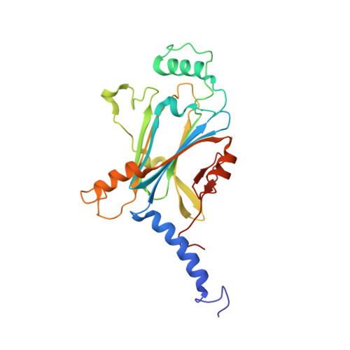
5GQM

Crystal structure of in cellulo Wild Type Cypovirus Polyhedra


Experimental Data Snapshot

  • Method: X-RAY DIFFRACTION
  • Resolution: 1.68 Å
  • R-Value Free: 0.196 
  • R-Value Work: 0.148 
  • R-Value Observed: 0.152 

Starting Model: experimental
View more details

wwPDB Validation   3D Report Full Report


Ligand Structure Quality Assessment 


This is version 1.4 of the entry. See complete history


Literature

Crystal Engineering of Self-Assembled Porous Protein Materials in Living Cells

Abe, S.Tabe, H.Ijiri, H.Yamashita, K.Hirata, K.Atsumi, K.Shimoi, T.Akai, M.Mori, H.Kitagawa, S.Ueno, T.

(2017) ACS Nano 11: 2410-2419

  • DOI: https://doi.org/10.1021/acsnano.6b06099
  • Primary Citation of Related Structures:  
    5GQI, 5GQJ, 5GQK, 5GQL, 5GQM, 5GQN

  • PubMed Abstract: 

    Crystalline porous materials have been investigated for development of important applications in molecular storage, separations, and catalysis. The potential of protein crystals is increasing as they become better understood. Protein crystals have been regarded as porous materials because they present highly ordered 3D arrangements of protein molecules with high porosity and wide range of pore sizes. However, it remains difficult to functionalize protein crystals in living cells. Here, we report that polyhedra, a natural crystalline protein assembly of polyhedrin monomer (PhM) produced in insect cells infected by cypovirus, can be engineered to extend porous networks by deleting selected amino acid residues located on the intermolecular contact region of PhM. The adsorption rates and quantities of fluorescent dyes stored within the mutant crystals are increased relative to those of the wild-type polyhedra crystal (WTPhC) under both in vitro and in vivo conditions. These results provide a strategy for designing self-assembled protein materials with applications in molecular recognition and storage of exogenous substances in living cell as well as an entry point for development of bioorthogonal chemistry and in vivo crystal structure analysis.


  • Organizational Affiliation

    School of Life Science and Technology, Tokyo Institute of Technology , Nagatsuta-cho, Midori-ku, Yokohama 226-8501, Japan.


Macromolecules
Find similar proteins by:  (by identity cutoff)  |  3D Structure
Entity ID: 1
MoleculeChains Sequence LengthOrganismDetailsImage
Polyhedrin248Bombyx mori cypovirus 1Mutation(s): 1 
UniProt
Find proteins for P11041 (Bombyx mori cytoplasmic polyhedrosis virus)
Explore P11041 
Go to UniProtKB:  P11041
Entity Groups  
Sequence Clusters30% Identity50% Identity70% Identity90% Identity95% Identity100% Identity
UniProt GroupP11041
Sequence Annotations
Expand
  • Reference Sequence
Small Molecules
Ligands 5 Unique
IDChains Name / Formula / InChI Key2D Diagram3D Interactions
GTP
Query on GTP

Download Ideal Coordinates CCD File 
E [auth A]GUANOSINE-5'-TRIPHOSPHATE
C10 H16 N5 O14 P3
XKMLYUALXHKNFT-UUOKFMHZSA-N
ATP
Query on ATP

Download Ideal Coordinates CCD File 
F [auth A]ADENOSINE-5'-TRIPHOSPHATE
C10 H16 N5 O13 P3
ZKHQWZAMYRWXGA-KQYNXXCUSA-N
CTP
Query on CTP

Download Ideal Coordinates CCD File 
G [auth A]CYTIDINE-5'-TRIPHOSPHATE
C9 H16 N3 O14 P3
PCDQPRRSZKQHHS-XVFCMESISA-N
CL
Query on CL

Download Ideal Coordinates CCD File 
B [auth A]CHLORIDE ION
Cl
VEXZGXHMUGYJMC-UHFFFAOYSA-M
MG
Query on MG

Download Ideal Coordinates CCD File 
C [auth A],
D [auth A]
MAGNESIUM ION
Mg
JLVVSXFLKOJNIY-UHFFFAOYSA-N
Experimental Data & Validation

Experimental Data

  • Method: X-RAY DIFFRACTION
  • Resolution: 1.68 Å
  • R-Value Free: 0.196 
  • R-Value Work: 0.148 
  • R-Value Observed: 0.152 
  • Space Group: I 2 3
Unit Cell:
Length ( Å )Angle ( ˚ )
a = 103.42α = 90
b = 103.42β = 90
c = 103.42γ = 90
Software Package:
Software NamePurpose
REFMACrefinement

Structure Validation

View Full Validation Report


Ligand Structure Quality Assessment 


Entry History 

Deposition Data

Revision History  (Full details and data files)

  • Version 1.0: 2017-02-15
    Type: Initial release
  • Version 1.1: 2017-04-12
    Changes: Database references
  • Version 1.2: 2020-02-26
    Changes: Data collection
  • Version 1.3: 2023-11-08
    Changes: Data collection, Database references, Derived calculations, Refinement description
  • Version 1.4: 2024-11-20
    Changes: Structure summary