


Crystal structure of ZapD, a positive regulator of Z-ring formation during bacterial cytokinesis
Lee, H.H., Choi, H., Min, K.J., Yoon, H.J., Ha, J.M.To be published.
Experimental Data Snapshot
Starting Model: experimental
View more details
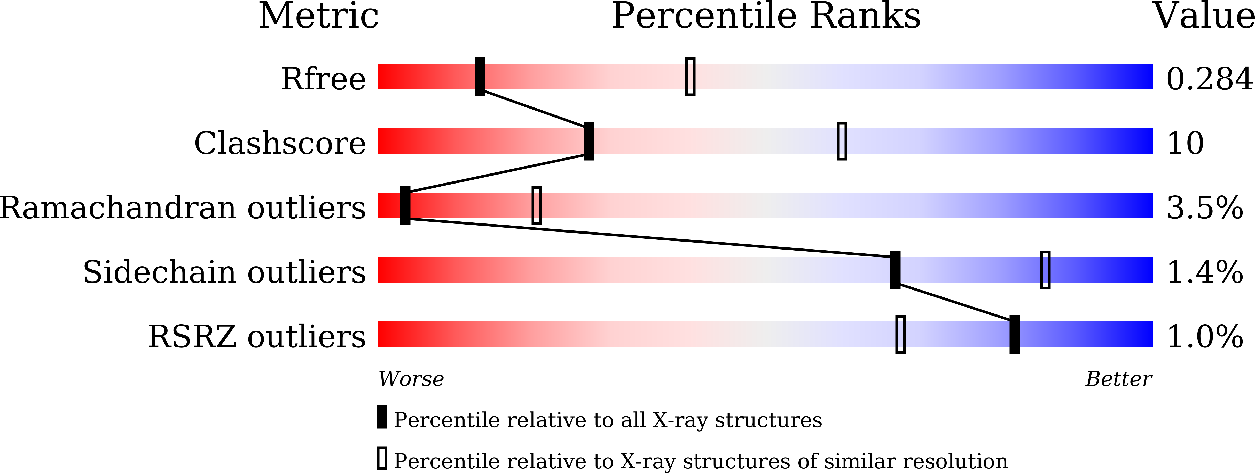
wwPDB Validation 3D Report Full Report
Entity ID: 1 | |||||
|---|---|---|---|---|---|
| Molecule | Chains | Sequence Length | Organism | Details | Image |
| Cell division protein ZapD | 246 | Escherichia coli K-12 | Mutation(s): 0 Gene Names: zapD, yacF, b0102, JW0099 |  | |
UniProt | |||||
Find proteins for P36680 (Escherichia coli (strain K12)) Explore P36680 Go to UniProtKB: P36680 | |||||
Entity Groups | |||||
| Sequence Clusters | 30% Identity50% Identity70% Identity90% Identity95% Identity100% Identity | ||||
| UniProt Group | P36680 | ||||
Sequence AnnotationsExpand | |||||
| |||||
| Ligands 1 Unique | |||||
|---|---|---|---|---|---|
| ID | Chains | Name / Formula / InChI Key | 2D Diagram | 3D Interactions | |
| SO4 Query on SO4 | C [auth A], D [auth A], E [auth A], F [auth B], G [auth B] | SULFATE ION O4 S QAOWNCQODCNURD-UHFFFAOYSA-L |  | ||
| Length ( Å ) | Angle ( ˚ ) |
|---|---|
| a = 109.49 | α = 90 |
| b = 109.49 | β = 90 |
| c = 106.659 | γ = 120 |
| Software Name | Purpose |
|---|---|
| REFMAC | refinement |
| HKL-2000 | data reduction |
| HKL-2000 | data scaling |
| MOLREP | phasing |


