5LZN

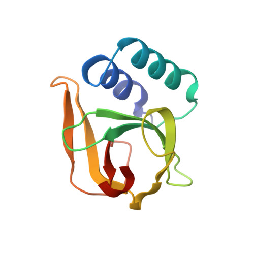
-TIP microtubule-binding domain


Experimental Data Snapshot

  • Method: X-RAY DIFFRACTION
  • Resolution: 1.40 Å
  • R-Value Free: 0.192 
  • R-Value Work: 0.162 
  • R-Value Observed: 0.164 

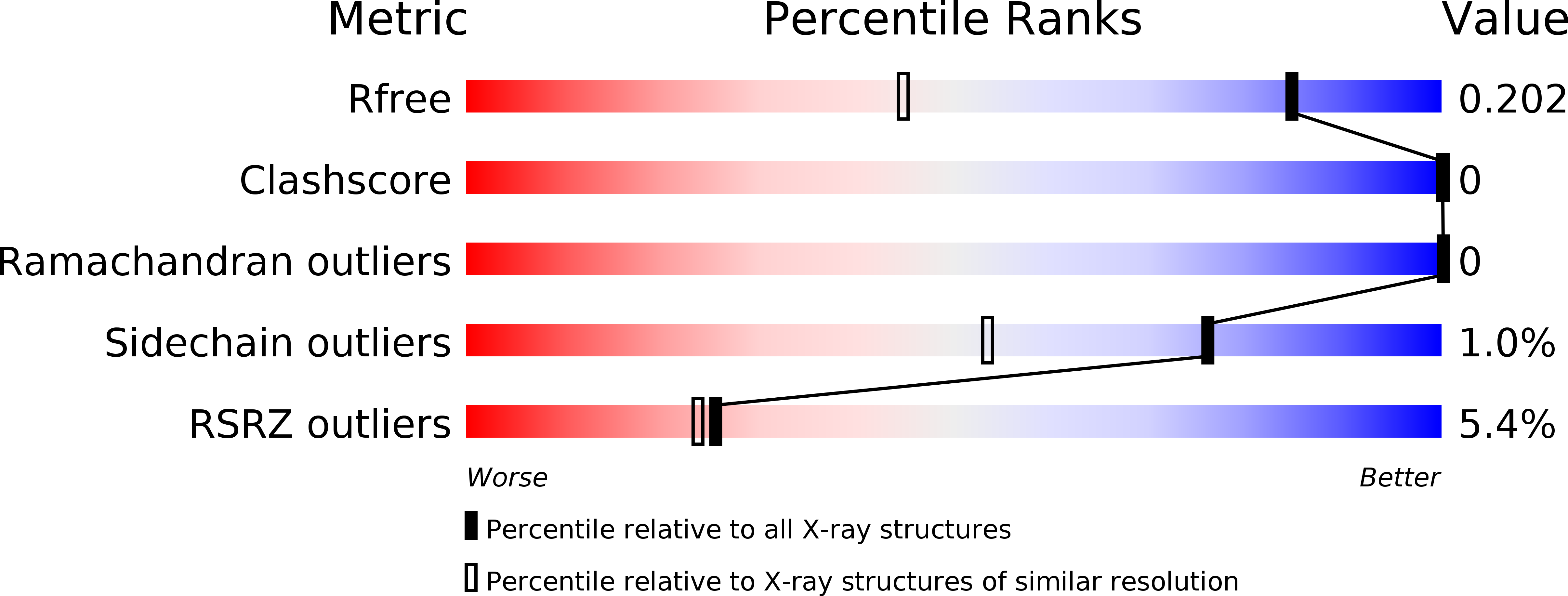
wwPDB Validation   3D Report Full Report


This is version 1.4 of the entry. See complete history


Literature

A structural model for microtubule minus-end recognition and protection by CAMSAP proteins.

Atherton, J.Jiang, K.Stangier, M.M.Luo, Y.Hua, S.Houben, K.van Hooff, J.J.E.Joseph, A.P.Scarabelli, G.Grant, B.J.Roberts, A.J.Topf, M.Steinmetz, M.O.Baldus, M.Moores, C.A.Akhmanova, A.

(2017) Nat Struct Mol Biol 24: 931-943

  • DOI: https://doi.org/10.1038/nsmb.3483
  • Primary Citation of Related Structures:  
    5LZN, 5M50, 5M54, 5M5C

  • PubMed Abstract: 

    CAMSAP and Patronin family members regulate microtubule minus-end stability and localization and thus organize noncentrosomal microtubule networks, which are essential for cell division, polarization and differentiation. Here, we found that the CAMSAP C-terminal CKK domain is widely present among eukaryotes and autonomously recognizes microtubule minus ends. Through a combination of structural approaches, we uncovered how mammalian CKK binds between two tubulin dimers at the interprotofilament interface on the outer microtubule surface. In vitro reconstitution assays combined with high-resolution fluorescence microscopy and cryo-electron tomography suggested that CKK preferentially associates with the transition zone between curved protofilaments and the regular microtubule lattice. We propose that minus-end-specific features of the interprotofilament interface at this site serve as the basis for CKK's minus-end preference. The steric clash between microtubule-bound CKK and kinesin motors explains how CKK protects microtubule minus ends against kinesin-13-induced depolymerization and thus controls the stability of free microtubule minus ends.


  • Organizational Affiliation

    Institute of Structural and Molecular Biology, Birkbeck, University of London, London, UK.


Macromolecules
Find similar proteins by:  (by identity cutoff)  |  3D Structure
Entity ID: 1
MoleculeChains Sequence LengthOrganismDetailsImage
Calmodulin-regulated spectrin-associated protein 3119Mus musculusMutation(s): 0 
Gene Names: Camsap3Kiaa1543
UniProt & NIH Common Fund Data Resources
Find proteins for Q80VC9 (Mus musculus)
Explore Q80VC9 
Go to UniProtKB:  Q80VC9
IMPC:  MGI:1916947
Entity Groups  
Sequence Clusters30% Identity50% Identity70% Identity90% Identity95% Identity100% Identity
UniProt GroupQ80VC9
Sequence Annotations
Expand
  • Reference Sequence
Experimental Data & Validation

Experimental Data

  • Method: X-RAY DIFFRACTION
  • Resolution: 1.40 Å
  • R-Value Free: 0.192 
  • R-Value Work: 0.162 
  • R-Value Observed: 0.164 
  • Space Group: I 4 2 2
Unit Cell:
Length ( Å )Angle ( ˚ )
a = 96.36α = 90
b = 96.36β = 90
c = 63.27γ = 90
Software Package:
Software NamePurpose
REFMACrefinement
XDSdata reduction
XSCALEdata scaling
PHASERphasing

Structure Validation

View Full Validation Report


Entry History & Funding Information

Deposition Data


Funding OrganizationLocationGrant Number
Swiss National Science FoundationSwitzerland--
SystemsX.chSwitzerland--

Revision History  (Full details and data files)

  • Version 1.0: 2017-10-04
    Type: Initial release
  • Version 1.1: 2017-10-18
    Changes: Database references
  • Version 1.2: 2017-11-15
    Changes: Database references
  • Version 1.3: 2019-10-16
    Changes: Data collection
  • Version 1.4: 2024-01-17
    Changes: Data collection, Database references, Refinement description