5MVV

Crystal structure of Plasmodium falciparum actin I- gelsolin segment 1 -CdATP complex


Experimental Data Snapshot

  • Method: X-RAY DIFFRACTION
  • Resolution: 1.40 Å
  • R-Value Free: 0.159 
  • R-Value Work: 0.129 
  • R-Value Observed: 0.129 

wwPDB Validation   3D Report Full Report


Ligand Structure Quality Assessment 


This is version 2.2 of the entry. See complete history


Literature

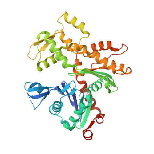
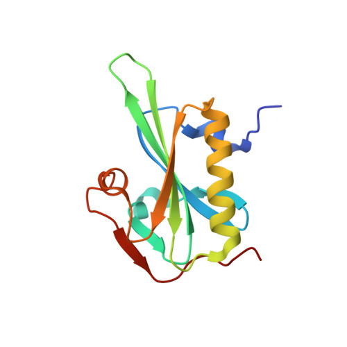
Rapid cadmium SAD phasing at the standard wavelength (1 angstrom ).

Panneerselvam, S.Kumpula, E.P.Kursula, I.Burkhardt, A.Meents, A.

(2017) Acta Crystallogr D Struct Biol 73: 581-590

  • DOI: https://doi.org/10.1107/S2059798317006970
  • Primary Citation of Related Structures:  
    5MVV, 5MYY

  • PubMed Abstract: 

    Cadmium ions can be effectively used to promote crystal growth and for experimental phasing. Here, the use of cadmium ions as a suitable anomalous scatterer at the standard wavelength of 1 Å is demonstrated. The structures of three different proteins were determined using cadmium single-wavelength anomalous dispersion (SAD) phasing. Owing to the strong anomalous signal, the structure of lysozyme could be automatically phased and built using a very low anomalous multiplicity (1.1) and low-completeness (77%) data set. Additionally, it is shown that cadmium ions can easily substitute divalent ions in ATP-divalent cation complexes. This property could be generally applied for phasing experiments of a wide range of nucleotide-binding proteins. Improvements in crystal growth and quality, good anomalous signal at standard wavelengths (i.e. no need to change photon energy) and rapid phasing and refinement using a single data set are benefits that should allow cadmium ions to be widely used for experimental phasing.


  • Organizational Affiliation

    Photon Science, DESY, Notkestrasse 85, 22607 Hamburg, Germany.


Macromolecules
Find similar proteins by:  (by identity cutoff)  |  3D Structure
Entity ID: 1
MoleculeChains Sequence LengthOrganismDetailsImage
Actin-1378Plasmodium falciparum 3D7Mutation(s): 0 
Gene Names: PFL2215w
UniProt
Find proteins for Q8I4X0 (Plasmodium falciparum (isolate 3D7))
Explore Q8I4X0 
Go to UniProtKB:  Q8I4X0
Entity Groups  
Sequence Clusters30% Identity50% Identity70% Identity90% Identity95% Identity100% Identity
UniProt GroupQ8I4X0
Sequence Annotations
Expand
  • Reference Sequence
Find similar proteins by:  (by identity cutoff)  |  3D Structure
Entity ID: 2
MoleculeChains Sequence LengthOrganismDetailsImage
GelsolinB [auth G]127Mus musculusMutation(s): 0 
Gene Names: GsnGsb
UniProt
Find proteins for P13020 (Mus musculus)
Explore P13020 
Go to UniProtKB:  P13020
Entity Groups  
Sequence Clusters30% Identity50% Identity70% Identity90% Identity95% Identity100% Identity
UniProt GroupP13020
Sequence Annotations
Expand
  • Reference Sequence
Small Molecules
Ligands 4 Unique
IDChains Name / Formula / InChI Key2D Diagram3D Interactions
ATP
Query on ATP

Download Ideal Coordinates CCD File 
C [auth A]ADENOSINE-5'-TRIPHOSPHATE
C10 H16 N5 O13 P3
ZKHQWZAMYRWXGA-KQYNXXCUSA-N
CD
Query on CD

Download Ideal Coordinates CCD File 
E [auth A],
F [auth A],
G [auth A]
CADMIUM ION
Cd
WLZRMCYVCSSEQC-UHFFFAOYSA-N
SCN
Query on SCN

Download Ideal Coordinates CCD File 
D [auth A]THIOCYANATE ION
C N S
ZMZDMBWJUHKJPS-UHFFFAOYSA-M
CA
Query on CA

Download Ideal Coordinates CCD File 
H [auth A],
I [auth G]
CALCIUM ION
Ca
BHPQYMZQTOCNFJ-UHFFFAOYSA-N
Experimental Data & Validation

Experimental Data

  • Method: X-RAY DIFFRACTION
  • Resolution: 1.40 Å
  • R-Value Free: 0.159 
  • R-Value Work: 0.129 
  • R-Value Observed: 0.129 
  • Space Group: P 21 21 2
Unit Cell:
Length ( Å )Angle ( ˚ )
a = 110.42α = 90
b = 68.92β = 90
c = 71.55γ = 90
Software Package:
Software NamePurpose
REFMACrefinement
XDSdata reduction
XSCALEdata scaling
SHELXDEphasing

Structure Validation

View Full Validation Report


Ligand Structure Quality Assessment 


Entry History 

Deposition Data

Revision History  (Full details and data files)

  • Version 1.0: 2017-07-12
    Type: Initial release
  • Version 2.0: 2017-07-19
    Changes: Atomic model
  • Version 2.1: 2017-08-02
    Changes: Database references
  • Version 2.2: 2019-10-16
    Changes: Data collection