5N6N

CRYSTAL STRUCTURE OF THE 14-3-3:NEUTRAL TREHALASE NTH1 COMPLEX


Experimental Data Snapshot

  • Method: X-RAY DIFFRACTION
  • Resolution: 2.29 Å
  • R-Value Free: 0.237 
  • R-Value Work: 0.184 
  • R-Value Observed: 0.186 

wwPDB Validation   3D Report Full Report


This is version 2.1 of the entry. See complete history


Literature

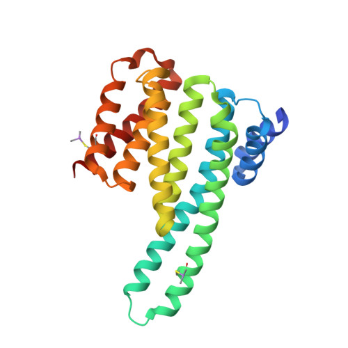
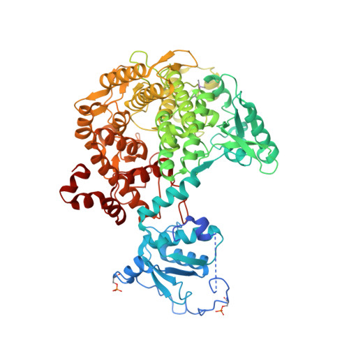
Molecular basis of the 14-3-3 protein-dependent activation of yeast neutral trehalase Nth1.

Alblova, M.Smidova, A.Docekal, V.Vesely, J.Herman, P.Obsilova, V.Obsil, T.

(2017) Proc Natl Acad Sci U S A 114: E9811-E9820

  • DOI: https://doi.org/10.1073/pnas.1714491114
  • Primary Citation of Related Structures:  
    5JTA, 5N6N, 5NIS

  • PubMed Abstract: 

    The 14-3-3 proteins, a family of highly conserved scaffolding proteins ubiquitously expressed in all eukaryotic cells, interact with and regulate the function of several hundreds of partner proteins. Yeast neutral trehalases (Nth), enzymes responsible for the hydrolysis of trehalose to glucose, compared with trehalases from other organisms, possess distinct structure and regulation involving phosphorylation at multiple sites followed by binding to the 14-3-3 protein. Here we report the crystal structures of yeast Nth1 and its complex with Bmh1 (yeast 14-3-3 isoform), which, together with mutational and fluorescence studies, indicate that the binding of Nth1 by 14-3-3 triggers Nth1's activity by enabling the proper 3D configuration of Nth1's catalytic and calcium-binding domains relative to each other, thus stabilizing the flexible part of the active site required for catalysis. The presented structure of the Bmh1:Nth1 complex highlights the ability of 14-3-3 to modulate the structure of a multidomain binding partner and to function as an allosteric effector. Furthermore, comparison of the Bmh1:Nth1 complex structure with those of 14-3-3:serotonin N -acetyltransferase and 14-3-3:heat shock protein beta-6 complexes revealed similarities in the 3D structures of bound partner proteins, suggesting the highly conserved nature of 14-3-3 affects the structures of many client proteins.


  • Organizational Affiliation

    Department of Structural Biology of Signaling Proteins, Division Biotechnology and Biomedicine Center of the Academy of Sciences and Charles University in Vestec (BIOCEV), Institute of Physiology, The Czech Academy of Sciences, Prague 14220, Czech Republic.


Macromolecules
Find similar proteins by:  (by identity cutoff)  |  3D Structure
Entity ID: 1
MoleculeChains Sequence LengthOrganismDetailsImage
Protein BMH1
A, B
240Saccharomyces cerevisiae S288CMutation(s): 0 
Gene Names: BMH1YER177W
UniProt
Find proteins for P29311 (Saccharomyces cerevisiae (strain ATCC 204508 / S288c))
Explore P29311 
Go to UniProtKB:  P29311
Entity Groups  
Sequence Clusters30% Identity50% Identity70% Identity90% Identity95% Identity100% Identity
UniProt GroupP29311
Sequence Annotations
Expand
  • Reference Sequence
Find similar proteins by:  (by identity cutoff)  |  3D Structure
Entity ID: 2
MoleculeChains Sequence LengthOrganismDetailsImage
Neutral trehalase756Saccharomyces cerevisiae S288CMutation(s): 0 
Gene Names: NTH1NTHYDR001CYD8119.07C
EC: 3.2.1.28
UniProt
Find proteins for P32356 (Saccharomyces cerevisiae (strain ATCC 204508 / S288c))
Explore P32356 
Go to UniProtKB:  P32356
Entity Groups  
Sequence Clusters30% Identity50% Identity70% Identity90% Identity95% Identity100% Identity
UniProt GroupP32356
Sequence Annotations
Expand
  • Reference Sequence
Oligosaccharides

Help

Entity ID: 3
MoleculeChains Length2D Diagram Glycosylation3D Interactions
beta-D-fructofuranose-(2-1)-alpha-D-glucopyranose
D, E, F
2N/A
Glycosylation Resources
GlyTouCan:  G05551OP
GlyCosmos:  G05551OP
Small Molecules
Ligands 1 Unique
IDChains Name / Formula / InChI Key2D Diagram3D Interactions
CA
Query on CA

Download Ideal Coordinates CCD File 
G [auth C]CALCIUM ION
Ca
BHPQYMZQTOCNFJ-UHFFFAOYSA-N
Modified Residues  2 Unique
IDChains TypeFormula2D DiagramParent
CAS
Query on CAS
A, B
L-PEPTIDE LINKINGC5 H12 As N O2 SCYS
SEP
Query on SEP
C
L-PEPTIDE LINKINGC3 H8 N O6 PSER
Biologically Interesting Molecules (External Reference) 1 Unique
Experimental Data & Validation

Experimental Data

  • Method: X-RAY DIFFRACTION
  • Resolution: 2.29 Å
  • R-Value Free: 0.237 
  • R-Value Work: 0.184 
  • R-Value Observed: 0.186 
  • Space Group: C 1 2 1
Unit Cell:
Length ( Å )Angle ( ˚ )
a = 160.805α = 90
b = 104.749β = 121.89
c = 105.638γ = 90
Software Package:
Software NamePurpose
PHENIXrefinement
XDSdata reduction
XDSdata scaling
MOLREPphasing

Structure Validation

View Full Validation Report


Entry History 

Deposition Data

Revision History  (Full details and data files)

  • Version 1.0: 2017-11-01
    Type: Initial release
  • Version 1.1: 2017-11-08
    Changes: Database references
  • Version 1.2: 2017-11-22
    Changes: Database references
  • Version 2.0: 2020-07-29
    Type: Remediation
    Reason: Carbohydrate remediation
    Changes: Atomic model, Data collection, Derived calculations, Non-polymer description, Structure summary
  • Version 2.1: 2024-01-17
    Changes: Data collection, Database references, Refinement description, Structure summary