5X2B | pdb_00005x2b

Crystal structure of mouse sulfotransferase SULT7A1 complexed with PAP


Experimental Data Snapshot

  • Method: X-RAY DIFFRACTION
  • Resolution: 2.08 Å
  • R-Value Free: 
    0.236 (Depositor), 0.220 (DCC) 
  • R-Value Work: 
    0.221 (Depositor), 0.200 (DCC) 
  • R-Value Observed: 
    0.222 (Depositor) 

wwPDB Validation   3D Report Full Report


Ligand Structure Quality Assessment 


This is version 1.2 of the entry. See complete history


Literature

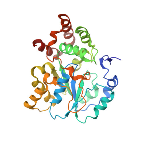
A new type of sulfation reaction: C -sulfonation for alpha , beta-unsaturated carbonyl groups by a novel sulfotransferase SULT7A1.

Kurogi, K.Sakakibara, Y.Hashiguchi, T.Kakuta, Y.Kanekiyo, M.Teramoto, T.Fukushima, T.Bamba, T.Matsumoto, J.Fukusaki, E.Kataoka, H.Suiko, M.

(2024) PNAS Nexus 3: pgae097-pgae097

  • DOI: https://doi.org/10.1093/pnasnexus/pgae097
  • Primary Citation of Related Structures:  
    5X2B

  • PubMed Abstract: 

    Cytosolic sulfotransferases (SULTs) are cytosolic enzymes that catalyze the transfer of sulfonate group to key endogenous compounds, altering the physiological functions of their substrates. SULT enzymes catalyze the O -sulfonation of hydroxy groups or N -sulfonation of amino groups of substrate compounds. In this study, we report the discovery of C -sulfonation of α,β-unsaturated carbonyl groups mediated by a new SULT enzyme, SULT7A1, and human SULT1C4. Enzymatic assays revealed that SULT7A1 is capable of transferring the sulfonate group from 3'-phosphoadenosine 5'-phosphosulfate to the α-carbon of α,β-unsaturated carbonyl-containing compounds, including cyclopentenone prostaglandins as representative endogenous substrates. Structural analyses of SULT7A1 suggest that the C -sulfonation reaction is catalyzed by a novel mechanism mediated by His and Cys residues in the active site. Ligand-activity assays demonstrated that sulfonated 15-deoxy prostaglandin J 2 exhibits antagonist activity against the prostaglandin receptor EP2 and the prostacyclin receptor IP. Modification of α,β-unsaturated carbonyl groups via the new prostaglandin-sulfonating enzyme, SULT7A1, may regulate the physiological function of prostaglandins in the gut. Discovery of C -sulfonation of α,β-unsaturated carbonyl groups will broaden the spectrum of potential substrates and physiological functions of SULTs.


  • Organizational Affiliation
    • Department of Biochemistry and Applied Biosciences, Faculty of Agriculture, University of Miyazaki, Miyazaki 889-2192, Japan.

Macromolecules
Find similar proteins by:  (by identity cutoff)  |  3D Structure
Entity ID: 1
MoleculeChains Sequence LengthOrganismDetailsImage
Sulfotransferase283Mus musculusMutation(s): 0 
Gene Names: Sult6b2Gm766
EC: 2.8.2
UniProt
Find proteins for B7ZWN4 (Mus musculus)
Explore B7ZWN4 
Go to UniProtKB:  B7ZWN4
Entity Groups  
Sequence Clusters30% Identity50% Identity70% Identity90% Identity95% Identity100% Identity
UniProt GroupB7ZWN4
Sequence Annotations
Expand
  • Reference Sequence
Find similar proteins by:  (by identity cutoff)  |  3D Structure
Entity ID: 2
MoleculeChains Sequence LengthOrganismDetailsImage
Sulfotransferase281Mus musculusMutation(s): 0 
Gene Names: Sult6b2Gm766
EC: 2.8.2
UniProt
Find proteins for B7ZWN4 (Mus musculus)
Explore B7ZWN4 
Go to UniProtKB:  B7ZWN4
Entity Groups  
Sequence Clusters30% Identity50% Identity70% Identity90% Identity95% Identity100% Identity
UniProt GroupB7ZWN4
Sequence Annotations
Expand
  • Reference Sequence
Find similar proteins by:  (by identity cutoff)  |  3D Structure
Entity ID: 3
MoleculeChains Sequence LengthOrganismDetailsImage
Sulfotransferase
F, G, H, J
282Mus musculusMutation(s): 0 
Gene Names: Sult6b2Gm766
EC: 2.8.2
UniProt
Find proteins for B7ZWN4 (Mus musculus)
Explore B7ZWN4 
Go to UniProtKB:  B7ZWN4
Entity Groups  
Sequence Clusters30% Identity50% Identity70% Identity90% Identity95% Identity100% Identity
UniProt GroupB7ZWN4
Sequence Annotations
Expand
  • Reference Sequence
Small Molecules
Ligands 3 Unique
IDChains Name / Formula / InChI Key2D Diagram3D Interactions
A3P
Query on A3P

Download Ideal Coordinates CCD File 
BA [auth F]
EA [auth G]
HA [auth H]
JA [auth I]
LA [auth J]
BA [auth F],
EA [auth G],
HA [auth H],
JA [auth I],
LA [auth J],
M [auth C],
O [auth A],
PA [auth K],
R [auth B],
SA [auth L],
V [auth D],
Z [auth E]
ADENOSINE-3'-5'-DIPHOSPHATE
C10 H15 N5 O10 P2
WHTCPDAXWFLDIH-KQYNXXCUSA-N
GOL
Query on GOL

Download Ideal Coordinates CCD File 
DA [auth F],
OA [auth J]
GLYCEROL
C3 H8 O3
PEDCQBHIVMGVHV-UHFFFAOYSA-N
CA
Query on CA

Download Ideal Coordinates CCD File 
AA [auth E]
CA [auth F]
FA [auth G]
GA [auth G]
IA [auth H]
AA [auth E],
CA [auth F],
FA [auth G],
GA [auth G],
IA [auth H],
KA [auth I],
MA [auth J],
N [auth C],
NA [auth J],
P [auth A],
Q [auth A],
QA [auth K],
RA [auth K],
S [auth B],
T [auth B],
TA [auth L],
U [auth B],
W [auth D],
X [auth D],
Y [auth D]
CALCIUM ION
Ca
BHPQYMZQTOCNFJ-UHFFFAOYSA-N
Experimental Data & Validation

Experimental Data

  • Method: X-RAY DIFFRACTION
  • Resolution: 2.08 Å
  • R-Value Free:  0.236 (Depositor), 0.220 (DCC) 
  • R-Value Work:  0.221 (Depositor), 0.200 (DCC) 
  • R-Value Observed: 0.222 (Depositor) 
Space Group: P 1 21 1
Unit Cell:
Length ( Å )Angle ( ˚ )
a = 166.174α = 90
b = 81.127β = 119.95
c = 166.18γ = 90
Software Package:
Software NamePurpose
REFMACrefinement
HKL-2000data reduction
HKL-2000data scaling
REFMACphasing

Structure Validation

View Full Validation Report


Ligand Structure Quality Assessment 


Entry History 

Deposition Data

Revision History  (Full details and data files)

  • Version 1.0: 2018-03-28
    Type: Initial release
  • Version 1.1: 2024-03-27
    Changes: Data collection, Database references, Derived calculations
  • Version 1.2: 2025-03-19
    Changes: Database references, Structure summary