


Crystal structure of adenine phosphoribosyltransferase from Yersinia pseudotuberculosis
Pavithra, G.C., Ramagopal, U.A.To be published.
Experimental Data Snapshot
Starting Model: experimental
View more details
Entity ID: 1 | |||||
|---|---|---|---|---|---|
| Molecule | Chains | Sequence Length | Organism | Details | Image |
| Adenine phosphoribosyltransferase | 187 | Yersinia pseudotuberculosis IP 32953 | Mutation(s): 0 Gene Names: YPTB0991 EC: 2.4.2.7 | 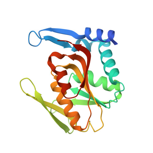 | |
UniProt | |||||
Find proteins for Q66DQ2 (Yersinia pseudotuberculosis serotype I (strain IP32953)) Explore Q66DQ2 Go to UniProtKB: Q66DQ2 | |||||
Entity Groups | |||||
| Sequence Clusters | 30% Identity50% Identity70% Identity90% Identity95% Identity100% Identity | ||||
| UniProt Group | Q66DQ2 | ||||
Sequence AnnotationsExpand | |||||
| |||||
| Ligands 2 Unique | |||||
|---|---|---|---|---|---|
| ID | Chains | Name / Formula / InChI Key | 2D Diagram | 3D Interactions | |
| ADE (Subject of Investigation/LOI) Query on ADE | B [auth A] | ADENINE C5 H5 N5 GFFGJBXGBJISGV-UHFFFAOYSA-N |  | ||
| NA Query on NA | C [auth A] | SODIUM ION Na FKNQFGJONOIPTF-UHFFFAOYSA-N |  | ||
| Length ( Å ) | Angle ( ˚ ) |
|---|---|
| a = 58.958 | α = 90 |
| b = 78.65 | β = 115.2 |
| c = 53.679 | γ = 90 |
| Software Name | Purpose |
|---|---|
| Aimless | data scaling |
| REFMAC | refinement |
| PDB_EXTRACT | data extraction |
| MOSFLM | data reduction |
| MOLREP | phasing |


