6CB3

Crystal structure of the L.Lactis YkoY riboswitch bound to cadmium


Experimental Data Snapshot

  • Method: X-RAY DIFFRACTION
  • Resolution: 1.89 Å
  • R-Value Free: 0.174 
  • R-Value Work: 0.140 
  • R-Value Observed: 0.147 

wwPDB Validation   3D Report Full Report


Ligand Structure Quality Assessment 


This is version 1.3 of the entry. See complete history


Literature

Convergent Use of Heptacoordination for Cation Selectivity by RNA and Protein Metalloregulators.

Bachas, S.T.Ferre-D'Amare, A.R.

(2018) Cell Chem Biol 25: 962-973.e5

  • DOI: https://doi.org/10.1016/j.chembiol.2018.04.016
  • Primary Citation of Related Structures:  
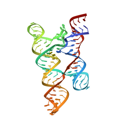
    6CB3, 6CC1, 6CC3

  • PubMed Abstract: 

    The large yybP-ykoY family of bacterial riboswitches is broadly distributed phylogenetically. Previously, these gene-regulatory RNAs were proposed to respond to Mn 2+ . X-ray crystallography revealed a binuclear cation-binding pocket. This comprises one hexacoordinate site, with six oxygen ligands, which preorganizes the second, with five oxygen and one nitrogen ligands. The relatively soft nitrogen ligand was proposed to confer affinity for Mn 2+ , but how this excludes other soft cations remained enigmatic. By subjecting representative yybP-ykoY riboswitches to diverse cations in vitro, we now find that these RNAs exhibit limited transition metal ion selectivity. Among the cations tested, Cd 2+ and Mn 2+ bind most tightly, and comparison of three new Cd 2+ -bound crystal structures suggests that these riboswitches achieve selectivity by enforcing heptacoordination (favored by high-spin Cd 2+ and Mn 2+ , but otherwise uncommon) in the softer site. Remarkably, the Cd 2+ - and Mn 2+ -selective bacterial transcription factor MntR also uses heptacoordination within a binuclear site to achieve selectivity.


  • Organizational Affiliation

    Biochemistry and Biophysics Center, National Heart, Lung and Blood Institute, Bethesda, MD 20892, USA.


Macromolecules
Find similar nucleic acids by:  (by identity cutoff)  |  3D Structure
Entity ID: 1
MoleculeChains LengthOrganismImage
RNA (99-MER)
A, B
101Lactococcus lactis
Sequence Annotations
Expand
  • Reference Sequence
Small Molecules
Ligands 4 Unique
IDChains Name / Formula / InChI Key2D Diagram3D Interactions
GTP
Query on GTP

Download Ideal Coordinates CCD File 
AB [auth B],
U [auth A]
GUANOSINE-5'-TRIPHOSPHATE
C10 H16 N5 O14 P3
XKMLYUALXHKNFT-UUOKFMHZSA-N
BA
Query on BA

Download Ideal Coordinates CCD File 
AA [auth A]
BA [auth A]
BB [auth B]
CB [auth B]
DB [auth B]
AA [auth A],
BA [auth A],
BB [auth B],
CB [auth B],
DB [auth B],
EB [auth B],
FB [auth B],
GB [auth B],
V [auth A],
W [auth A],
X [auth A],
Y [auth A],
Z [auth A]
BARIUM ION
Ba
XDFCIPNJCBUZJN-UHFFFAOYSA-N
CD
Query on CD

Download Ideal Coordinates CCD File 
C [auth A]
D [auth A]
E [auth A]
F [auth A]
G [auth A]
C [auth A],
D [auth A],
E [auth A],
F [auth A],
G [auth A],
GA [auth B],
H [auth A],
HA [auth B],
I [auth A],
IA [auth B],
J [auth A],
JA [auth B],
K [auth A],
KA [auth B],
L [auth A],
LA [auth B],
M [auth A],
MA [auth B],
N [auth A],
NA [auth B],
O [auth A],
OA [auth B],
P [auth A],
PA [auth B],
Q [auth A],
QA [auth B],
R [auth A],
RA [auth B],
S [auth A],
SA [auth B],
T [auth A],
TA [auth B],
UA [auth B],
VA [auth B],
WA [auth B],
XA [auth B],
YA [auth B],
ZA [auth B]
CADMIUM ION
Cd
WLZRMCYVCSSEQC-UHFFFAOYSA-N
MG
Query on MG

Download Ideal Coordinates CCD File 
CA [auth A]
DA [auth A]
EA [auth A]
FA [auth A]
HB [auth B]
CA [auth A],
DA [auth A],
EA [auth A],
FA [auth A],
HB [auth B],
IB [auth B]
MAGNESIUM ION
Mg
JLVVSXFLKOJNIY-UHFFFAOYSA-N
Experimental Data & Validation

Experimental Data

  • Method: X-RAY DIFFRACTION
  • Resolution: 1.89 Å
  • R-Value Free: 0.174 
  • R-Value Work: 0.140 
  • R-Value Observed: 0.147 
  • Space Group: P 31
Unit Cell:
Length ( Å )Angle ( ˚ )
a = 73.141α = 90
b = 73.141β = 90
c = 117.61γ = 120
Software Package:
Software NamePurpose
PHENIXrefinement
HKL-2000data reduction
HKL-2000data scaling
PHASERphasing

Structure Validation

View Full Validation Report


Ligand Structure Quality Assessment 


Entry History 

Deposition Data

Revision History  (Full details and data files)

  • Version 1.0: 2018-06-13
    Type: Initial release
  • Version 1.1: 2018-08-29
    Changes: Data collection, Database references
  • Version 1.2: 2019-12-04
    Changes: Author supporting evidence
  • Version 1.3: 2023-10-04
    Changes: Data collection, Database references, Derived calculations, Refinement description