6CEY

Aminoglycoside Phosphotransferase (2'')-Ia in complex with GMPPNP, Magnesium, and Lividomycin moieties


Experimental Data Snapshot

  • Method: X-RAY DIFFRACTION
  • Resolution: 2.40 Å
  • R-Value Free: 0.216 
  • R-Value Work: 0.171 
  • R-Value Observed: 0.174 

wwPDB Validation   3D Report Full Report


Ligand Structure Quality Assessment 


This is version 1.5 of the entry. See complete history


Literature

Plasticity of Aminoglycoside Binding to Antibiotic Kinase APH(2′′)-Ia.

Caldwell, S.J.Berghuis, A.M.

(2018) Antimicrob Agents Chemother 62

  • DOI: https://doi.org/10.1128/AAC.00202-18
  • Primary Citation of Related Structures:  
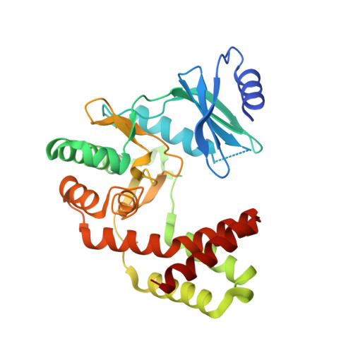
    6C5U, 6CAV, 6CEY, 6CGD, 6CGG, 6CH4

  • PubMed Abstract: 

    The APH(2″)-Ia aminoglycoside resistance enzyme forms the C-terminal domain of the bifunctional AAC(6')-Ie/APH(2″)-Ia enzyme and confers high-level resistance to natural 4,6-disubstituted aminoglycosides. In addition, reports have suggested that the enzyme can phosphorylate 4,5-disubstituted compounds and aminoglycosides with substitutions at the N1 position. Previously determined structures of the enzyme with bound aminoglycosides have not indicated how these noncanonical substrates may bind and be modified by the enzyme. We carried out crystallographic studies to directly observe the interactions of these compounds with the aminoglycoside binding site and to probe the means by which these noncanonical substrates interact with the enzyme. We find that APH(2″)-Ia maintains a preferred mode of binding aminoglycosides by using the conserved neamine rings when possible, with flexibility that allows it to accommodate additional rings. However, if this binding mode is made impossible because of additional substitutions to the standard 4,5- or 4,6-disubstituted aminoglycoside architecture, as in lividomycin A or the N1-substituted aminoglycosides, it is still possible for these aminoglycosides to bind to the antibiotic binding site by using alternate binding modes, which explains the low rates of noncanonical phosphorylation activities seen in enzyme assays. Furthermore, structural studies of a clinically observed arbekacin-resistant mutant of APH(2″)-Ia revealed an altered aminoglycoside binding site that can stabilize an alternative binding mode for N1-substituted aminoglycosides. This mutation may alter and expand the aminoglycoside resistance spectrum of the wild-type enzyme in response to newly developed aminoglycosides.


  • Organizational Affiliation

    Department of Biochemistry, McGill University, Montreal, Canada.


Macromolecules
Find similar proteins by:  (by identity cutoff)  |  3D Structure
Entity ID: 1
MoleculeChains Sequence LengthOrganismDetailsImage
Bifunctional AAC/APH
A, B, C, D
305Staphylococcus aureusMutation(s): 0 
Gene Names: aacA-aphDR015VRA0030
EC: 2.3.1 (PDB Primary Data), 2.7.1.190 (PDB Primary Data)
UniProt
Find proteins for P0A0C1 (Staphylococcus aureus)
Explore P0A0C1 
Go to UniProtKB:  P0A0C1
Entity Groups  
Sequence Clusters30% Identity50% Identity70% Identity90% Identity95% Identity100% Identity
UniProt GroupP0A0C1
Sequence Annotations
Expand
  • Reference Sequence
Small Molecules
Ligands 4 Unique
IDChains Name / Formula / InChI Key2D Diagram3D Interactions
LIV
Query on LIV

Download Ideal Coordinates CCD File 
S [auth D](2R,3S,4S,5S,6R)-2-((2S,3S,4R,5R,6R)-5-AMINO-2-(AMINOMETHYL)-6-((2R,3S,4R,5S)-5-((1R,2R,3S,5R,6S)-3,5-DIAMINO-2-((2S,3R ,5S,6R)-3-AMINO-5-HYDROXY-6-(HYDROXYMETHYL)-TETRAHYDRO-2H-PYRAN-2-YLOXY)-6-HYDROXYCYCLOHEXYLOXY)-4-HYDROXY-2-(HYDROXYMET HYL)-TETRAHYDROFURAN-3-YLOXY)-4-HYDROXY-TETRAHYDRO-2H-PYRAN-3-YLOXY)-6-(HYDROXYMETHYL)-TETRAHYDRO-2H-PYRAN-3,4,5-TRIOL
C29 H55 N5 O18
DBLVDAUGBTYDFR-SWMBIRFSSA-N
GNP
Query on GNP

Download Ideal Coordinates CCD File 
E [auth A],
I [auth B],
M [auth C],
R [auth D]
PHOSPHOAMINOPHOSPHONIC ACID-GUANYLATE ESTER
C10 H17 N6 O13 P3
UQABYHGXWYXDTK-UUOKFMHZSA-N
CL
Query on CL

Download Ideal Coordinates CCD File 
H [auth A],
L [auth B],
P [auth C]
CHLORIDE ION
Cl
VEXZGXHMUGYJMC-UHFFFAOYSA-M
MG
Query on MG

Download Ideal Coordinates CCD File 
F [auth A]
G [auth A]
J [auth B]
K [auth B]
N [auth C]
F [auth A],
G [auth A],
J [auth B],
K [auth B],
N [auth C],
O [auth C],
Q [auth C],
T [auth D],
U [auth D]
MAGNESIUM ION
Mg
JLVVSXFLKOJNIY-UHFFFAOYSA-N
Experimental Data & Validation

Experimental Data

  • Method: X-RAY DIFFRACTION
  • Resolution: 2.40 Å
  • R-Value Free: 0.216 
  • R-Value Work: 0.171 
  • R-Value Observed: 0.174 
  • Space Group: P 1 21 1
Unit Cell:
Length ( Å )Angle ( ˚ )
a = 90.25α = 90
b = 100.24β = 105.17
c = 94.01γ = 90
Software Package:
Software NamePurpose
REFMACrefinement
iMOSFLMdata reduction
Aimlessdata scaling
Cootmodel building

Structure Validation

View Full Validation Report


Ligand Structure Quality Assessment 


Entry History & Funding Information

Deposition Data


Funding OrganizationLocationGrant Number
Canadian Institutes of Health Research (CIHR)CanadaMOP-13107

Revision History  (Full details and data files)

  • Version 1.0: 2018-04-11
    Type: Initial release
  • Version 1.1: 2018-05-02
    Changes: Data collection, Database references
  • Version 1.2: 2018-07-04
    Changes: Data collection, Database references
  • Version 1.3: 2019-02-20
    Changes: Author supporting evidence, Data collection
  • Version 1.4: 2020-01-08
    Changes: Author supporting evidence
  • Version 1.5: 2023-10-04
    Changes: Data collection, Database references, Derived calculations, Refinement description, Structure summary