6CIN

Crystal structure of pyruvate:ferredoxin oxidoreductase from Moorella thermoacetica


Experimental Data Snapshot

  • Method: X-RAY DIFFRACTION
  • Resolution: 2.60 Å
  • R-Value Free: 0.227 
  • R-Value Work: 0.195 
  • R-Value Observed: 0.197 

wwPDB Validation   3D Report Full Report


Ligand Structure Quality Assessment 


This is version 1.6 of the entry. See complete history


Literature

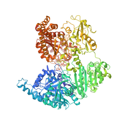
Binding site for coenzyme A revealed in the structure of pyruvate:ferredoxin oxidoreductase fromMoorella thermoacetica.

Chen, P.Y.Aman, H.Can, M.Ragsdale, S.W.Drennan, C.L.

(2018) Proc Natl Acad Sci U S A 115: 3846-3851

  • DOI: https://doi.org/10.1073/pnas.1722329115
  • Primary Citation of Related Structures:  
    6CIN, 6CIO, 6CIP, 6CIQ

  • PubMed Abstract: 

    Pyruvate:ferredoxin oxidoreductase (PFOR) is a microbial enzyme that uses thiamine pyrophosphate (TPP), three [4Fe-4S] clusters, and coenzyme A (CoA) in the reversible oxidation of pyruvate to generate acetyl-CoA and carbon dioxide. The two electrons that are generated as a result of pyruvate decarboxylation are used in the reduction of low potential ferredoxins, which provide reducing equivalents for central metabolism, including the Wood-Ljungdahl pathway. PFOR is a member of the 2-oxoacid:ferredoxin oxidoreductase (OFOR) superfamily, which plays major roles in both microbial redox reactions and carbon dioxide fixation. Here, we present a set of crystallographic snapshots of the best-studied member of this superfamily, the PFOR from Moorella thermoacetica ( Mt PFOR). These snapshots include the native structure, those of lactyl-TPP and acetyl-TPP reaction intermediates, and the first of an OFOR with CoA bound. These structural data reveal the binding site of CoA as domain III, the function of which in OFORs was previously unknown, and establish sequence motifs for CoA binding in the OFOR superfamily. Mt PFOR structures further show that domain III undergoes a conformational change upon CoA binding that seals off the active site and positions the thiolate of CoA directly adjacent to the TPP cofactor. These structural findings provide a molecular basis for the experimental observation that CoA binding accelerates catalysis by 10 5 -fold.


  • Organizational Affiliation

    Department of Chemistry, Massachusetts Institute of Technology, Cambridge, MA 02139.


Macromolecules
Find similar proteins by:  (by identity cutoff)  |  3D Structure
Entity ID: 1
MoleculeChains Sequence LengthOrganismDetailsImage
PYRUVATE-FERREDOXIN OXIDOREDUCTASE
A, B, C, D, E
A, B, C, D, E, F
1,171Moorella thermoacetica ATCC 39073Mutation(s): 0 
Gene Names: Moth_0064
EC: 1.2.7
UniProt
Find proteins for Q2RMD6 (Moorella thermoacetica (strain ATCC 39073 / JCM 9320))
Explore Q2RMD6 
Go to UniProtKB:  Q2RMD6
Entity Groups  
Sequence Clusters30% Identity50% Identity70% Identity90% Identity95% Identity100% Identity
UniProt GroupQ2RMD6
Sequence Annotations
Expand
  • Reference Sequence
Small Molecules
Ligands 4 Unique
IDChains Name / Formula / InChI Key2D Diagram3D Interactions
TPP
Query on TPP

Download Ideal Coordinates CCD File 
CA [auth D]
IA [auth E]
J [auth A]
OA [auth F]
P [auth B]
CA [auth D],
IA [auth E],
J [auth A],
OA [auth F],
P [auth B],
W [auth C]
THIAMINE DIPHOSPHATE
C12 H19 N4 O7 P2 S
AYEKOFBPNLCAJY-UHFFFAOYSA-O
SF4
Query on SF4

Download Ideal Coordinates CCD File 
AA [auth D]
BA [auth D]
FA [auth E]
G [auth A]
GA [auth E]
AA [auth D],
BA [auth D],
FA [auth E],
G [auth A],
GA [auth E],
H [auth A],
HA [auth E],
I [auth A],
LA [auth F],
M [auth B],
MA [auth F],
N [auth B],
NA [auth F],
O [auth B],
T [auth C],
U [auth C],
V [auth C],
Z [auth D]
IRON/SULFUR CLUSTER
Fe4 S4
LJBDFODJNLIPKO-UHFFFAOYSA-N
SO4
Query on SO4

Download Ideal Coordinates CCD File 
EA [auth D]
KA [auth E]
L [auth A]
QA [auth F]
R [auth B]
EA [auth D],
KA [auth E],
L [auth A],
QA [auth F],
R [auth B],
S [auth B],
Y [auth C]
SULFATE ION
O4 S
QAOWNCQODCNURD-UHFFFAOYSA-L
MG
Query on MG

Download Ideal Coordinates CCD File 
DA [auth D]
JA [auth E]
K [auth A]
PA [auth F]
Q [auth B]
DA [auth D],
JA [auth E],
K [auth A],
PA [auth F],
Q [auth B],
X [auth C]
MAGNESIUM ION
Mg
JLVVSXFLKOJNIY-UHFFFAOYSA-N
Experimental Data & Validation

Experimental Data

  • Method: X-RAY DIFFRACTION
  • Resolution: 2.60 Å
  • R-Value Free: 0.227 
  • R-Value Work: 0.195 
  • R-Value Observed: 0.197 
  • Space Group: C 1 2 1
Unit Cell:
Length ( Å )Angle ( ˚ )
a = 340.611α = 90
b = 106.634β = 109.31
c = 239.079γ = 90
Software Package:
Software NamePurpose
PHENIXrefinement
HKL-2000data reduction
HKL-2000data scaling
PHENIXphasing

Structure Validation

View Full Validation Report


Ligand Structure Quality Assessment 


Entry History & Funding Information

Deposition Data


Funding OrganizationLocationGrant Number
National Institutes of Health/National Institute of General Medical Sciences (NIH/NIGMS)United StatesGM069857
National Institutes of Health/National Institute of General Medical Sciences (NIH/NIGMS)United StatesGM39451

Revision History  (Full details and data files)

  • Version 1.0: 2018-03-28
    Type: Initial release
  • Version 1.1: 2018-04-11
    Changes: Data collection, Database references
  • Version 1.2: 2018-04-25
    Changes: Data collection, Database references
  • Version 1.3: 2018-10-03
    Changes: Data collection, Database references
  • Version 1.4: 2019-02-20
    Changes: Author supporting evidence, Data collection
  • Version 1.5: 2020-01-01
    Changes: Author supporting evidence
  • Version 1.6: 2023-10-04
    Changes: Data collection, Database references, Derived calculations, Refinement description