6CS6

CryoEM structure of human enterovirus D68 A-particle (pH 5.5 and room temperature)

  • Classification: VIRUS
  • Organism(s): enterovirus D68
  • Mutation(s): No 

  • Deposited: 2018-03-19 Released: 2018-12-19 
  • Deposition Author(s): Liu, Y., Rossmann, M.G.
  • Funding Organization(s): National Institutes of Health/National Human Genome Research Institute (NIH/NHGRI)

Experimental Data Snapshot

  • Method: ELECTRON MICROSCOPY
  • Resolution: 3.25 Å
  • Aggregation State: PARTICLE 
  • Reconstruction Method: SINGLE PARTICLE 

wwPDB Validation   3D Report Full Report


This is version 1.4 of the entry. See complete history


Literature

Molecular basis for the acid-initiated uncoating of human enterovirus D68.

Liu, Y.Sheng, J.van Vliet, A.L.W.Buda, G.van Kuppeveld, F.J.M.Rossmann, M.G.

(2018) Proc Natl Acad Sci U S A 115: E12209-E12217

  • DOI: https://doi.org/10.1073/pnas.1803347115
  • Primary Citation of Related Structures:  
    6CRP, 6CRR, 6CRS, 6CRU, 6CS3, 6CS4, 6CS5, 6CS6, 6CSA, 6CSG, 6CSH, 6MZI

  • PubMed Abstract: 

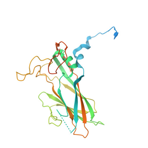
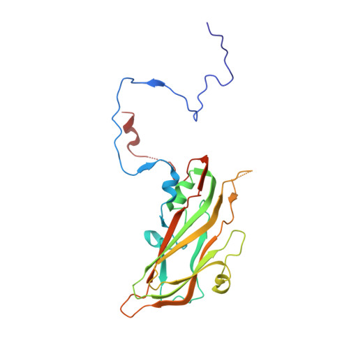
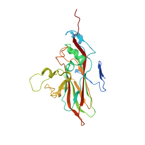
    Enterovirus D68 (EV-D68) belongs to a group of enteroviruses that contain a single positive-sense RNA genome surrounded by an icosahedral capsid. Like common cold viruses, EV-D68 mainly causes respiratory infections and is acid-labile. The molecular mechanism by which the acid-sensitive EV-D68 virions uncoat and deliver their genome into a host cell is unknown. Using cryoelectron microscopy (cryo-EM), we have determined the structures of the full native virion and an uncoating intermediate [the A (altered) particle] of EV-D68 at 2.2- and 2.7-Å resolution, respectively. These structures showed that acid treatment of EV-D68 leads to particle expansion, externalization of the viral protein VP1 N termini from the capsid interior, and formation of pores around the icosahedral twofold axes through which the viral RNA can exit. Moreover, because of the low stability of EV-D68, cryo-EM analyses of a mixed population of particles at neutral pH and following acid treatment demonstrated the involvement of multiple structural intermediates during virus uncoating. Among these, a previously undescribed state, the expanded 1 ("E1") particle, shows a majority of internal regions (e.g., the VP1 N termini) to be ordered as in the full native virion. Thus, the E1 particle acts as an intermediate in the transition from full native virions to A particles. Together, the present work delineates the pathway of EV-D68 uncoating and provides the molecular basis for the acid lability of EV-D68 and of the related common cold viruses.


  • Organizational Affiliation

    Department of Biological Sciences, Purdue University, West Lafayette, IN 47907.


Macromolecules
Find similar proteins by:  (by identity cutoff)  |  3D Structure
Entity ID: 1
MoleculeChains Sequence LengthOrganismDetailsImage
viral protein 1297enterovirus D68Mutation(s): 0 
UniProt
Find proteins for A0A097BW12 (Human enterovirus D68)
Explore A0A097BW12 
Go to UniProtKB:  A0A097BW12
Entity Groups  
Sequence Clusters30% Identity50% Identity70% Identity90% Identity95% Identity100% Identity
UniProt GroupA0A097BW12
Sequence Annotations
Expand
  • Reference Sequence
Find similar proteins by:  (by identity cutoff)  |  3D Structure
Entity ID: 2
MoleculeChains Sequence LengthOrganismDetailsImage
viral protein 3247enterovirus D68Mutation(s): 0 
UniProt
Find proteins for A0A097BW12 (Human enterovirus D68)
Explore A0A097BW12 
Go to UniProtKB:  A0A097BW12
Entity Groups  
Sequence Clusters30% Identity50% Identity70% Identity90% Identity95% Identity100% Identity
UniProt GroupA0A097BW12
Sequence Annotations
Expand
  • Reference Sequence
Find similar proteins by:  (by identity cutoff)  |  3D Structure
Entity ID: 3
MoleculeChains Sequence LengthOrganismDetailsImage
viral protein 2248enterovirus D68Mutation(s): 0 
UniProt
Find proteins for A0A097BW12 (Human enterovirus D68)
Explore A0A097BW12 
Go to UniProtKB:  A0A097BW12
Entity Groups  
Sequence Clusters30% Identity50% Identity70% Identity90% Identity95% Identity100% Identity
UniProt GroupA0A097BW12
Sequence Annotations
Expand
  • Reference Sequence
Experimental Data & Validation

Experimental Data

  • Method: ELECTRON MICROSCOPY
  • Resolution: 3.25 Å
  • Aggregation State: PARTICLE 
  • Reconstruction Method: SINGLE PARTICLE 
EM Software:
TaskSoftware PackageVersion
MODEL REFINEMENTPHENIX
MODEL REFINEMENTCoot
MODEL REFINEMENTREFMAC
RECONSTRUCTIONjspr

Structure Validation

View Full Validation Report


Entry History & Funding Information

Deposition Data


Funding OrganizationLocationGrant Number
National Institutes of Health/National Human Genome Research Institute (NIH/NHGRI)United StatesAI011219

Revision History  (Full details and data files)

  • Version 1.0: 2018-12-19
    Type: Initial release
  • Version 1.1: 2018-12-26
    Changes: Data collection, Database references
  • Version 1.2: 2019-01-09
    Changes: Data collection, Database references
  • Version 1.3: 2019-12-18
    Changes: Author supporting evidence
  • Version 1.4: 2024-03-13
    Changes: Data collection, Database references, Derived calculations