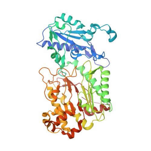
Structural basis for prolidase deficiency disease mechanisms.
Wilk, P., Uehlein, M., Piwowarczyk, R., Dobbek, H., Mueller, U., Weiss, M.S.(2018) FEBS J 285: 3422-3441
- PubMed: 30066404
- DOI: https://doi.org/10.1111/febs.14620
- Primary Citation of Related Structures:
5MBY, 5MBZ, 5MC0, 5MC1, 5MC2, 5MC3, 5MC4, 5MC5, 6H2P, 6H2Q - PubMed Abstract:
Prolidase is a metallopeptidase that cleaves iminodipeptides containing a proline (Pro) or hydroxyproline (Hyp) residue at their C-terminal end. The disease prolidase deficiency (PD) is a rare recessive human disorder characterized by reduced prolidase activity. PD manifests itself by a wide range of severe clinical symptoms, most commonly as skin ulceration, recurrent infections of the respiratory tract, and mental retardation. Several mutations in the PEPD gene have been identified that are responsible for the loss or the reduction of prolidase activity. In contrast, the structural basis of enzyme inactivation has so far remained elusive. In this study, we present high resolution crystal structures of a number of human prolidase (HsProl) variants, in which single amino acids are either substituted by others or deleted. The observed implications of the mutations on the three-dimensional structure of HsProl are reported and discussed and related to their enzymatic activity. The resulting structures may be divided into four groups depending on the presumed effect of the corresponding mutations on the reaction mechanism. The four possible inactivation mechanisms, which could be elucidated, are disruption of the catalytic Mn 2 (OH - )-center, introduction of chain disorder along with the displacement of important active site residues, rigidification of the active site, and flexibilization of the active site. All refined structure coordinates as well as the corresponding structure factor amplitudes have been deposited in the PDB under the accession numbers 5MBY, 5MBZ, 5MC0, 5MC1, 5MC2, 5MC3, 5MC4, 5MC5, 6H2P, 6H2Q.
Organizational Affiliation:
Helmholtz-Zentrum Berlin, Macromolecular Crystallography (HZB-MX), Germany.