6I9Z | pdb_00006i9z

urate oxidase under 65 bar of argon


Experimental Data Snapshot

  • Method: X-RAY DIFFRACTION
  • Resolution: 1.60 Å
  • R-Value Free: 
    0.183 (Depositor), 0.190 (DCC) 
  • R-Value Work: 
    0.156 (Depositor), 0.170 (DCC) 
  • R-Value Observed: 
    0.157 (Depositor) 

Starting Model: experimental
View more details

wwPDB Validation   3D Report Full Report


Ligand Structure Quality Assessment 


This is version 1.4 of the entry. See complete history


Literature

Comparative study of the effects of high hydrostatic pressure per se and high argon pressure on urate oxidase ligand stabilization.

Prange, T.Carpentier, P.Dhaussy, A.C.van der Linden, P.Girard, E.Colloc'h, N.

(2022) Acta Crystallogr D Struct Biol 78: 162-173

  • DOI: https://doi.org/10.1107/S2059798321012134
  • Primary Citation of Related Structures:  
    6I9X, 6I9Z, 6IA1, 6IA3, 6IA9, 7P0C, 7P0D, 7P0G, 7PUF, 7PWN, 7Q09

  • PubMed Abstract: 

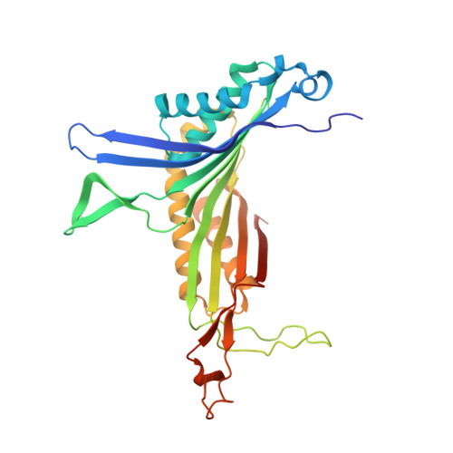
    The stability of the tetrameric enzyme urate oxidase in complex with excess of 8-azaxanthine was investigated either under high hydrostatic pressure per se or under a high pressure of argon. The active site is located at the interface of two subunits, and the catalytic activity is directly related to the integrity of the tetramer. This study demonstrates that applying pressure to a protein-ligand complex drives the thermodynamic equilibrium towards ligand saturation of the complex, revealing a new binding site. A transient dimeric intermediate that occurs during the pressure-induced dissociation process was characterized under argon pressure and excited substates of the enzyme that occur during the catalytic cycle can be trapped by pressure. Comparison of the different structures under pressure infers an allosteric role of the internal hydrophobic cavity in which argon is bound, since this cavity provides the necessary flexibility for the active site to function.


  • Organizational Affiliation

    CiTCoM UMR 8038, CNRS, Université de Paris, Faculté de Pharmacie, 4 Avenue de l'Observatoire, 75006 Paris, France.


Macromolecules
Find similar proteins by:  (by identity cutoff)  |  3D Structure
Entity ID: 1
MoleculeChains Sequence LengthOrganismDetailsImage
Uricase302Aspergillus flavusMutation(s): 0 
Gene Names: uaZuox
EC: 1.7.3.3
UniProt
Find proteins for Q00511 (Aspergillus flavus)
Explore Q00511 
Go to UniProtKB:  Q00511
Entity Groups  
Sequence Clusters30% Identity50% Identity70% Identity90% Identity95% Identity100% Identity
UniProt GroupQ00511
Sequence Annotations
Expand
  • Reference Sequence
Experimental Data & Validation

Experimental Data

  • Method: X-RAY DIFFRACTION
  • Resolution: 1.60 Å
  • R-Value Free:  0.183 (Depositor), 0.190 (DCC) 
  • R-Value Work:  0.156 (Depositor), 0.170 (DCC) 
  • R-Value Observed: 0.157 (Depositor) 
Space Group: I 2 2 2
Unit Cell:
Length ( Å )Angle ( ˚ )
a = 80.323α = 90
b = 96.121β = 90
c = 105.412γ = 90
Software Package:
Software NamePurpose
REFMACrefinement
HKL-2000data reduction
SCALEPACKdata scaling
FFTphasing

Structure Validation

View Full Validation Report


Ligand Structure Quality Assessment 


Entry History 

Deposition Data

Revision History  (Full details and data files)

  • Version 1.0: 2019-12-18
    Type: Initial release
  • Version 1.1: 2022-02-09
    Changes: Database references, Derived calculations
  • Version 1.2: 2024-01-24
    Changes: Data collection, Refinement description
  • Version 1.3: 2024-11-06
    Changes: Structure summary
  • Version 1.4: 2025-07-23
    Changes: Database references