


Endothiapepsin in complex with ligand 69
Magari, F., Heine, A., Konstantinidou, M., Sutanto, F., Haupenthal, J., Jumde, R.V., Unver, M.Y., Camacho, C.J., Hirsch, A.K.H., Doemling, A., Klebe, G.To be published.
Experimental Data Snapshot
Entity ID: 1 | |||||
|---|---|---|---|---|---|
| Molecule | Chains | Sequence Length | Organism | Details | Image |
| Endothiapepsin | 330 | Cryphonectria parasitica | Mutation(s): 0 EC: 3.4.23.22 | 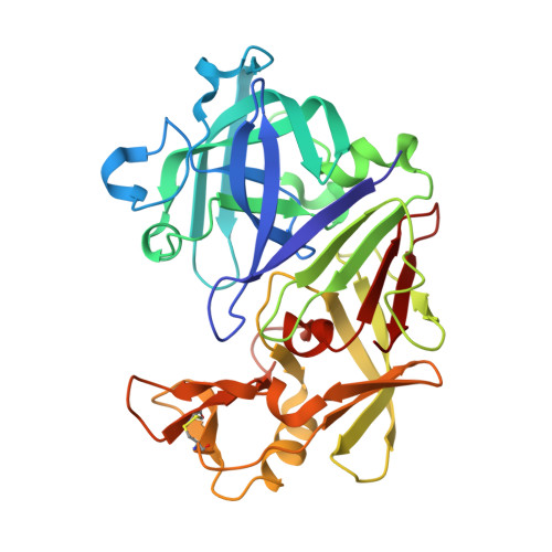 | |
UniProt | |||||
Find proteins for P11838 (Cryphonectria parasitica) Explore P11838 Go to UniProtKB: P11838 | |||||
Entity Groups | |||||
| Sequence Clusters | 30% Identity50% Identity70% Identity90% Identity95% Identity100% Identity | ||||
| UniProt Group | P11838 | ||||
Sequence AnnotationsExpand | |||||
| |||||
| Ligands 2 Unique | |||||
|---|---|---|---|---|---|
| ID | Chains | Name / Formula / InChI Key | 2D Diagram | 3D Interactions | |
| L7K (Subject of Investigation/LOI) Query on L7K | C [auth A] | [(~{R})-cyclohexyl-[1-(2-phenylethyl)-1,2,3,4-tetrazol-5-yl]methyl]diazane C16 H24 N6 RYAQXBFFYZPWMF-OAHLLOKOSA-N |  | ||
| GOL Query on GOL | B [auth A] | GLYCEROL C3 H8 O3 PEDCQBHIVMGVHV-UHFFFAOYSA-N |  | ||
| Length ( Å ) | Angle ( ˚ ) |
|---|---|
| a = 45.283 | α = 90 |
| b = 73.04 | β = 109.378 |
| c = 52.878 | γ = 90 |
| Software Name | Purpose |
|---|---|
| PHENIX | refinement |
| XDS | data reduction |
| XDS | data scaling |
| PHASER | phasing |


