6TB0 | pdb_00006tb0

Crystal structure of thermostable omega transaminase 4-fold mutant from Pseudomonas jessenii


Experimental Data Snapshot

  • Method: X-RAY DIFFRACTION
  • Resolution: 1.95 Å
  • R-Value Free: 
    0.169 (Depositor), 0.180 (DCC) 
  • R-Value Work: 
    0.140 (Depositor), 0.150 (DCC) 
  • R-Value Observed: 
    0.142 (Depositor) 

Starting Model: experimental
View more details

wwPDB Validation   3D Report Full Report


Ligand Structure Quality Assessment 


This is version 1.2 of the entry. See complete history


Literature

Robust omega-Transaminases by Computational Stabilization of the Subunit Interface.

Meng, Q.Capra, N.Palacio, C.M.Lanfranchi, E.Otzen, M.van Schie, L.Z.Rozeboom, H.J.Thunnissen, A.W.H.Wijma, H.J.Janssen, D.B.

(2020) ACS Catal 10: 2915-2928

  • DOI: https://doi.org/10.1021/acscatal.9b05223
  • Primary Citation of Related Structures:  
    6TB0, 6TB1

  • PubMed Abstract: 

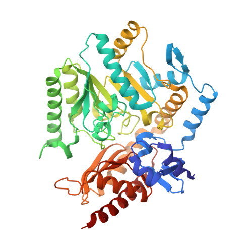
    Transaminases are attractive catalysts for the production of enantiopure amines. However, the poor stability of these enzymes often limits their application in biocatalysis. Here, we used a framework for enzyme stability engineering by computational library design (FRESCO) to stabilize the homodimeric PLP fold type I ω-transaminase from Pseudomonas jessenii . A large number of surface-located point mutations and mutations predicted to stabilize the subunit interface were examined. Experimental screening revealed that 10 surface mutations out of 172 tested were indeed stabilizing (6% success), whereas testing 34 interface mutations gave 19 hits (56% success). Both the extent of stabilization and the spatial distribution of stabilizing mutations showed that the subunit interface was critical for stability. After mutations were combined, 2 very stable variants with 4 and 6 mutations were obtained, which in comparison to wild type ( T m app = 62 °C) displayed T m app values of 80 and 85 °C, respectively. These two variants were also 5-fold more active at their optimum temperatures and tolerated high concentrations of isopropylamine and cosolvents. This allowed conversion of 100 mM acetophenone to ( S )-1-phenylethylamine (>99% enantiomeric excess) with high yield (92%, in comparison to 24% with the wild-type transaminase). Crystal structures mostly confirmed the expected structural changes and revealed that the most stabilizing mutation, I154V, featured a rarely described stabilization mechanism: namely, removal of steric strain. The results show that computational interface redesign can be a rapid and powerful strategy for transaminase stabilization.


  • Organizational Affiliation
    • Biotransformation and Biocatalysis, Groningen Biomolecular Sciences and Biotechnology Institute (GBB), University of Groningen, Nijenborgh 4, 9747 AG Groningen, The Netherlands.

Macromolecules
Find similar proteins by:  (by identity cutoff)  |  3D Structure
Entity ID: 1
MoleculeChains Sequence LengthOrganismDetailsImage
Aspartate aminotransferase family protein
A, B
453Pseudomonas sp.Mutation(s): 4 
Gene Names: CMK94_18730DIU04_17820
Entity Groups  
Sequence Clusters30% Identity50% Identity70% Identity90% Identity95% Identity100% Identity
Sequence Annotations
Expand
  • Reference Sequence
Small Molecules
Ligands 3 Unique
IDChains Name / Formula / InChI Key2D Diagram3D Interactions
PLP
Query on PLP

Download Ideal Coordinates CCD File 
C [auth A],
F [auth B]
PYRIDOXAL-5'-PHOSPHATE
C8 H10 N O6 P
NGVDGCNFYWLIFO-UHFFFAOYSA-N
SIN
Query on SIN

Download Ideal Coordinates CCD File 
G [auth B]SUCCINIC ACID
C4 H6 O4
KDYFGRWQOYBRFD-UHFFFAOYSA-N
GOL
Query on GOL

Download Ideal Coordinates CCD File 
D [auth A],
E [auth A],
H [auth B],
I [auth B]
GLYCEROL
C3 H8 O3
PEDCQBHIVMGVHV-UHFFFAOYSA-N
Experimental Data & Validation

Experimental Data

  • Method: X-RAY DIFFRACTION
  • Resolution: 1.95 Å
  • R-Value Free:  0.169 (Depositor), 0.180 (DCC) 
  • R-Value Work:  0.140 (Depositor), 0.150 (DCC) 
  • R-Value Observed: 0.142 (Depositor) 
Space Group: P 43
Unit Cell:
Length ( Å )Angle ( ˚ )
a = 98.07α = 90
b = 98.07β = 90
c = 119.31γ = 90
Software Package:
Software NamePurpose
REFMACrefinement
Aimlessdata scaling
PDB_EXTRACTdata extraction
iMOSFLMdata reduction
REFMACphasing

Structure Validation

View Full Validation Report


Ligand Structure Quality Assessment 


Entry History & Funding Information

Deposition Data


Funding OrganizationLocationGrant Number
European UnionNetherlands722610

Revision History  (Full details and data files)

  • Version 1.0: 2020-07-15
    Type: Initial release
  • Version 1.1: 2020-09-30
    Changes: Database references
  • Version 1.2: 2024-01-24
    Changes: Data collection, Database references, Refinement description