7PB6 | pdb_00007pb6

Structure of LPMO domain of colonization factor GbpA from Vibrio cholerae in the presence of potassium


Experimental Data Snapshot

  • Method: X-RAY DIFFRACTION
  • Resolution: 1.50 Å
  • R-Value Free: 
    0.192 (Depositor), 0.190 (DCC) 
  • R-Value Work: 
    0.164 (Depositor), 0.160 (DCC) 
  • R-Value Observed: 
    0.166 (Depositor) 

Starting Model: experimental
View more details

wwPDB Validation   3D Report Full Report


This is version 1.3 of the entry. See complete history


Literature

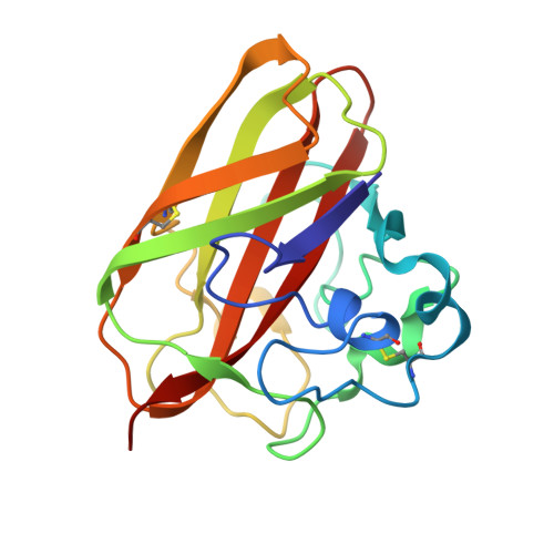
Calcium-binding site in AA10 LPMO from Vibrio cholerae suggests modulating effects during environmental survival and infection.

Montserrat-Canals, M.Bjerregaard-Andersen, K.Sorensen, H.V.Kommedal, E.Cordara, G.Vaaje-Kolstad, G.Krengel, U.

(2024) QRB Discov 5: e12-e12

  • DOI: https://doi.org/10.1017/qrd.2024.14
  • Primary Citation of Related Structures:  
    7PB6, 7PB7

  • PubMed Abstract: 

    Despite major efforts toward its eradication, cholera remains a major health threat and economic burden in many low- and middle-income countries. Between outbreaks, the bacterium responsible for the disease, Vibrio cholerae , survives in aquatic environmental reservoirs, where it commonly forms biofilms, for example, on zooplankton. N -acetyl glucosamine-binding protein A (GbpA) is an adhesin that binds to the chitinaceous surface of zooplankton and breaks its dense crystalline packing thanks to its lytic polysaccharide monooxygenase (LPMO) activity, which provides V. cholerae with nutrients. In addition, GbpA is an important colonization factor associated with bacterial pathogenicity, allowing the binding to mucins in the host intestine. Here, we report the discovery of a cation-binding site in proximity of the GbpA active site, which allows Ca 2+ , Mg 2+ , or K + binding close to its carbohydrate-binding surface. In addition to the X-ray crystal structures of cation-LPMO complexes (to 1.5 Å resolution), we explored how the presence of ions affects the stability and activity of the protein. Calcium and magnesium ions were found to bind to GbpA specifically, with calcium ions - abundant in natural sources of chitin - having the strongest effect on protein stability. When the cation-binding site was rendered non-functional, a decrease in activity was observed, highlighting the importance of the structural elements stabilized by calcium. Our findings suggest a cation-binding site specific to GbpA and related LPMOs that may fine-tune binding and activity for its substrates during environmental survival and host infection.


  • Organizational Affiliation
    • Centre for Molecular Medicine Norway, University of Oslo, NO-0318 Oslo, Norway.

Macromolecules
Find similar proteins by:  (by identity cutoff)  |  3D Structure
Entity ID: 1
MoleculeChains Sequence LengthOrganismDetailsImage
GlcNAc-binding protein A
A, B
180Vibrio cholerae O1Mutation(s): 0 
Gene Names: gbpAVC_A0811
UniProt
Find proteins for Q9KLD5 (Vibrio cholerae serotype O1 (strain ATCC 39315 / El Tor Inaba N16961))
Explore Q9KLD5 
Go to UniProtKB:  Q9KLD5
Entity Groups  
Sequence Clusters30% Identity50% Identity70% Identity90% Identity95% Identity100% Identity
UniProt GroupQ9KLD5
Sequence Annotations
Expand
  • Reference Sequence
Small Molecules
Ligands 3 Unique
IDChains Name / Formula / InChI Key2D Diagram3D Interactions
CU
Query on CU

Download Ideal Coordinates CCD File 
C [auth A],
L [auth B]
COPPER (II) ION
Cu
JPVYNHNXODAKFH-UHFFFAOYSA-N
SCN
Query on SCN

Download Ideal Coordinates CCD File 
I [auth A],
J [auth A],
K [auth A],
O [auth B],
P [auth B]
THIOCYANATE ION
C N S
ZMZDMBWJUHKJPS-UHFFFAOYSA-M
K
Query on K

Download Ideal Coordinates CCD File 
D [auth A]
E [auth A]
F [auth A]
G [auth A]
H [auth A]
D [auth A],
E [auth A],
F [auth A],
G [auth A],
H [auth A],
M [auth B],
N [auth B]
POTASSIUM ION
K
NPYPAHLBTDXSSS-UHFFFAOYSA-N
Experimental Data & Validation

Experimental Data

  • Method: X-RAY DIFFRACTION
  • Resolution: 1.50 Å
  • R-Value Free:  0.192 (Depositor), 0.190 (DCC) 
  • R-Value Work:  0.164 (Depositor), 0.160 (DCC) 
  • R-Value Observed: 0.166 (Depositor) 
Space Group: P 43
Unit Cell:
Length ( Å )Angle ( ˚ )
a = 46.58α = 90
b = 46.58β = 90
c = 157.43γ = 90
Software Package:
Software NamePurpose
PHENIXrefinement
Cootmodel building
XDSdata reduction
XSCALEdata scaling
PHENIXphasing

Structure Validation

View Full Validation Report


Entry History & Funding Information

Deposition Data


Funding OrganizationLocationGrant Number
Norwegian Research CouncilNorway247730

Revision History  (Full details and data files)

  • Version 1.0: 2022-08-10
    Type: Initial release
  • Version 1.1: 2024-01-31
    Changes: Data collection, Refinement description
  • Version 1.2: 2024-11-20
    Changes: Structure summary
  • Version 1.3: 2025-02-19
    Changes: Database references