7X2B | pdb_00007x2b

Red fluorescent protein from Diadumene lineata


Experimental Data Snapshot

  • Method: X-RAY DIFFRACTION
  • Resolution: 1.63 Å
  • R-Value Free: 
    0.205 (Depositor), 0.210 (DCC) 
  • R-Value Work: 
    0.171 (Depositor), 0.180 (DCC) 

Starting Model: experimental
View more details

wwPDB Validation   3D Report Full Report


This is version 2.1 of the entry. See complete history


Literature

Cloning and structural basis of fluorescent protein color variants from identical species of sea anemone, Diadumene lineata.

Horiuchi, Y.Makabe, K.Laskaratou, D.Hatori, K.Sliwa, M.Mizuno, H.Hotta, J.I.

(2023) Photochem Photobiol Sci 22: 1591-1601

  • DOI: https://doi.org/10.1007/s43630-023-00399-0
  • Primary Citation of Related Structures:  
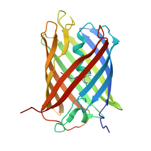
    7X2B

  • PubMed Abstract: 

    Diadumene lineata is a colorful sea anemone with orange stripe tissue of the body column and plain tentacles with red lines. We subjected Diadumene lineata to expression cloning and obtained genes encoding orange (OFP: DiLiFP561) and red fluorescent proteins (RFPs: DiLiFP570 and DiLiFP571). These proteins formed obligatory tetramers. All three proteins showed bright fluorescence with the brightness of 58.3 mM -1 ·cm -1 (DiLiFP561), 43.9 mM -1 ·cm -1 (DiLiFP570), and 31.2 mM -1 ·cm -1 (DiLiFP571), which were equivalent to that of commonly used red fluorescent proteins. Amplitude-weighted average fluorescence lifetimes of DiLiFP561, DiLiFP570 and DiLiFP571 were determined as 3.7, 3.6 and 3.0 ns. We determined a crystal structure of DiLiFP570 at 1.63 Å resolution. The crystal structure of DiLiFP570 revealed that the chromophore has an extended π-conjugated structure similar to that of DsRed. Most of the amino acid residues surrounding the chromophore were common between DiLiFP570 and DiLiFP561, except M159 of DiLiFP570 (Lysine in DiLiFP561), which is located close to the chromophore hydroxyl group. Interestingly, a similar K-to-M substitution has been reported in a red-shifted variant of DsRed (mRFP1). It is a striking observation that the naturally evolved color-change variants are consistent with the mutation induced via protein engineering processes. The newly cloned proteins are promising as orange and red fluorescent markers for imaging with long fluorescence lifetime.


  • Organizational Affiliation
    • Graduate School of Science and Engineering, Yamagata University, 4-3-16 Jonan, Yonezawa, Yamagata, 992-8510, Japan.

Macromolecules
Find similar proteins by:  (by identity cutoff)  |  3D Structure
Entity ID: 1
MoleculeChains Sequence LengthOrganismDetailsImage
Red fluorescent protein221Diadumene lineataMutation(s): 0 
UniProt
Find proteins for A0A9Y2YAB8 (Diadumene lineata)
Explore A0A9Y2YAB8 
Go to UniProtKB:  A0A9Y2YAB8
Entity Groups  
Sequence Clusters30% Identity50% Identity70% Identity90% Identity95% Identity100% Identity
UniProt GroupA0A9Y2YAB8
Sequence Annotations
Expand
  • Reference Sequence
Small Molecules
Ligands 1 Unique
IDChains Name / Formula / InChI Key2D Diagram3D Interactions
SO4
Query on SO4

Download Ideal Coordinates CCD File 
B [auth A]SULFATE ION
O4 S
QAOWNCQODCNURD-UHFFFAOYSA-L
Modified Residues  1 Unique
IDChains TypeFormula2D DiagramParent
NRQ
Query on NRQ
A
L-PEPTIDE LINKINGC16 H17 N3 O4 SMET, TYR, GLY
Experimental Data & Validation

Experimental Data

  • Method: X-RAY DIFFRACTION
  • Resolution: 1.63 Å
  • R-Value Free:  0.205 (Depositor), 0.210 (DCC) 
  • R-Value Work:  0.171 (Depositor), 0.180 (DCC) 
Space Group: I 2 2 2
Unit Cell:
Length ( Å )Angle ( ˚ )
a = 46.929α = 90
b = 75.394β = 90
c = 118.955γ = 90
Software Package:
Software NamePurpose
REFMACrefinement
XDSdata reduction
PHENIXrefinement

Structure Validation

View Full Validation Report


Entry History & Funding Information

Deposition Data


Funding OrganizationLocationGrant Number
Not funded--

Revision History  (Full details and data files)

  • Version 1.0: 2023-03-01
    Type: Initial release
  • Version 1.1: 2023-11-29
    Changes: Data collection, Refinement description
  • Version 2.0: 2024-01-31
    Changes: Advisory, Atomic model, Data collection, Derived calculations, Structure summary
  • Version 2.1: 2024-03-13
    Changes: Database references