7Y6E | pdb_00007y6e

Crystal structure of sDscam FNIII23 domains, isoform Beta2v6


Experimental Data Snapshot

  • Method: X-RAY DIFFRACTION
  • Resolution: 3.03 Å
  • R-Value Free: 
    0.272 (Depositor), 0.270 (DCC) 
  • R-Value Work: 
    0.234 (Depositor), 0.230 (DCC) 
  • R-Value Observed: 
    0.236 (Depositor) 

Starting Model: in silico
View more details

wwPDB Validation   3D Report Full Report


This is version 1.1 of the entry. See complete history


Literature

Structural basis for the self-recognition of sDSCAM in Chelicerata.

Cheng, J.Yu, Y.Wang, X.Zheng, X.Liu, T.Hu, D.Jin, Y.Lai, Y.Fu, T.M.Chen, Q.

(2023) Nat Commun 14: 2522-2522

  • DOI: https://doi.org/10.1038/s41467-023-38205-1
  • Primary Citation of Related Structures:  
    7Y4X, 7Y54, 7Y5J, 7Y5R, 7Y6E, 7Y6O, 7Y73, 7Y8H, 7Y8I, 7Y8S, 7Y95, 7Y9A

  • PubMed Abstract: 

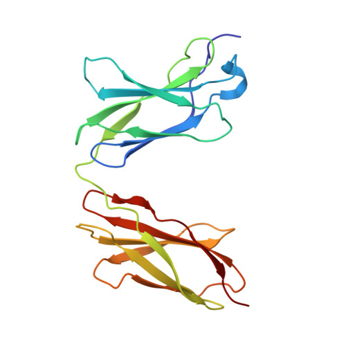
    To create a functional neural circuit, neurons develop a molecular identity to discriminate self from non-self. The invertebrate Dscam family and vertebrate Pcdh family are implicated in determining synaptic specificity. Recently identified in Chelicerata, a shortened Dscam (sDscam) has been shown to resemble the isoform-generating characters of both Dscam and Pcdh and represent an evolutionary transition. Here we presented the molecular details of sDscam self-recognition via both trans and cis interactions using X-ray crystallographic data and functional assays. Based on our results, we proposed a molecular zipper model for the assemblies of sDscam to mediate cell-cell recognition. In this model, sDscam utilized FNIII domain to form side-by-side interactions with neighboring molecules in the same cell while established hand-in-hand interactions via Ig1 domain with molecules from another cell around. Together, our study provided a framework for understanding the assembly, recognition, and evolution of sDscam.


  • Organizational Affiliation
    • National Clinical Research Center for Geriatrics, West China Hospital, State Key Laboratory of Biotherapy and Collaborative Innovation Center of Biotherapy, Sichuan University, 610041, Chengdu, China.

Macromolecules
Find similar proteins by:  (by identity cutoff)  |  3D Structure
Entity ID: 1
MoleculeChains Sequence LengthOrganismDetailsImage
Dscam
A, B, C, D, E
A, B, C, D, E, F, G
202ChelicerataMutation(s): 0 
Gene Names: Dscam
UniProt
Find proteins for A0A161FW14 (Olivierus martensii)
Explore A0A161FW14 
Go to UniProtKB:  A0A161FW14
Entity Groups  
Sequence Clusters30% Identity50% Identity70% Identity90% Identity95% Identity100% Identity
UniProt GroupA0A161FW14
Sequence Annotations
Expand
  • Reference Sequence
Experimental Data & Validation

Experimental Data

  • Method: X-RAY DIFFRACTION
  • Resolution: 3.03 Å
  • R-Value Free:  0.272 (Depositor), 0.270 (DCC) 
  • R-Value Work:  0.234 (Depositor), 0.230 (DCC) 
  • R-Value Observed: 0.236 (Depositor) 
Space Group: C 1 2 1
Unit Cell:
Length ( Å )Angle ( ˚ )
a = 307.021α = 90
b = 56.219β = 105.38
c = 94.837γ = 90
Software Package:
Software NamePurpose
PHENIXrefinement
HKL-2000data scaling
HKL-3000data reduction
PHASERphasing

Structure Validation

View Full Validation Report


Entry History & Funding Information

Deposition Data


Funding OrganizationLocationGrant Number
National Natural Science Foundation of China (NSFC)China--

Revision History  (Full details and data files)

  • Version 1.0: 2023-05-24
    Type: Initial release
  • Version 1.1: 2024-05-29
    Changes: Data collection