Neutron crystallography and quantum chemical analysis of bilin reductase PcyA mutants reveal substrate and catalytic residue protonation states.
Joutsuka, T., Nanasawa, R., Igarashi, K., Horie, K., Sugishima, M., Hagiwara, Y., Wada, K., Fukuyama, K., Yano, N., Mori, S., Ostermann, A., Kusaka, K., Unno, M.(2022) J Biological Chem 299: 102763-102763
- PubMed: 36463961
- DOI: https://doi.org/10.1016/j.jbc.2022.102763
- Primary Citation of Related Structures:
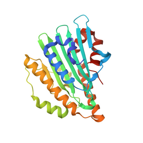
7YK9, 7YKB - PubMed Abstract:
PcyA, a ferredoxin-dependent bilin pigment reductase, catalyzes the site-specific reduction of the two vinyl groups of biliverdin (BV), producing phycocyanobilin. Previous neutron crystallography detected both the neutral BV and its protonated form (BVH + ) in the wildtype (WT) PcyA-BV complex, and a nearby catalytic residue Asp105 was found to have two conformations (protonated and deprotonated). Semiempirical calculations have suggested that the protonation states of BV are reflected in the absorption spectrum of the WT PcyA-BV complex. In the previously determined absorption spectra of the PcyA D105N and I86D mutants, complexed with BV, a peak at 730 nm, observed in the WT, disappeared and increased, respectively. Here, we performed neutron crystallography and quantum chemical analysis of the D105N-BV and I86D-BV complexes to determine the protonation states of BV and the surrounding residues and study the correlation between the absorption spectra and protonation states around BV. Neutron structures elucidated that BV in the D105N mutant is in a neutral state, whereas that in the I86D mutant is dominantly in a protonated state. Glu76 and His88 showed different hydrogen bonding with surrounding residues compared with WT PcyA, further explaining why D105N and I86D have much lower activities for phycocyanobilin synthesis than the WT PcyA. Our quantum mechanics/molecular mechanics calculations of the absorption spectra showed that the spectral change in D105N arises from Glu76 deprotonation, consistent with the neutron structure. Collectively, our findings reveal more mechanistic details of bilin pigment biosynthesis.
- Graduate School of Science and Engineering, Ibaraki University, Hitachi, Ibaraki, Japan; Frontier Research Center for Applied Atomic Sciences, Ibaraki University, Naka-Tokai, Ibaraki, Japan. Electronic address: tatsuya.joutsuka.joe@vc.ibaraki.ac.jp.
Organizational Affiliation: