7Z43

Influenza B polymerase with Pol II pSer5 CTD peptide mimic bound in site 1B and 2B


Experimental Data Snapshot

  • Method: X-RAY DIFFRACTION
  • Resolution: 3.12 Å
  • R-Value Free: 0.272 
  • R-Value Work: 0.240 

wwPDB Validation   3D Report Full Report


Ligand Structure Quality Assessment 


This is version 1.2 of the entry. See complete history


Literature

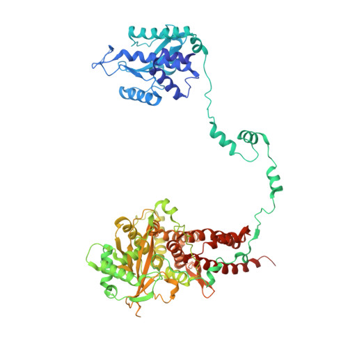
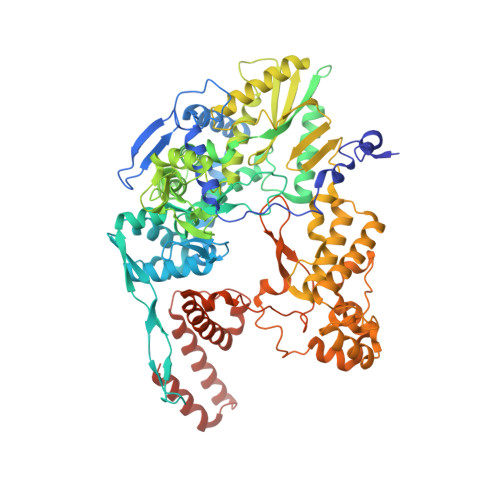
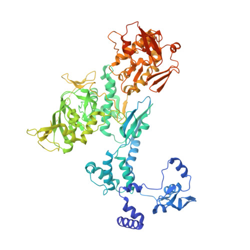
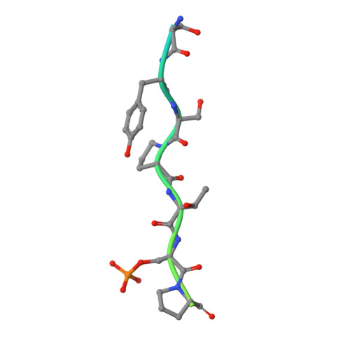
Type B and type A influenza polymerases have evolved distinct binding interfaces to recruit the RNA polymerase II CTD.

Krischuns, T.Isel, C.Drncova, P.Lukarska, M.Pflug, A.Paisant, S.Navratil, V.Cusack, S.Naffakh, N.

(2022) PLoS Pathog 18: e1010328-e1010328

  • DOI: https://doi.org/10.1371/journal.ppat.1010328
  • Primary Citation of Related Structures:  
    7Z42, 7Z43, 7Z4O

  • PubMed Abstract: 

    During annual influenza epidemics, influenza B viruses (IBVs) co-circulate with influenza A viruses (IAVs), can become predominant and cause severe morbidity and mortality. Phylogenetic analyses suggest that IAVs (primarily avian viruses) and IBVs (primarily human viruses) have diverged over long time scales. Identifying their common and distinctive features is an effective approach to increase knowledge about the molecular details of influenza infection. The virus-encoded RNA-dependent RNA polymerases (FluPolB and FluPolA) are PB1-PB2-PA heterotrimers that perform transcription and replication of the viral genome in the nucleus of infected cells. Initiation of viral mRNA synthesis requires a direct association of FluPol with the host RNA polymerase II (RNAP II), in particular the repetitive C-terminal domain (CTD) of the major RNAP II subunit, to enable "cap-snatching" whereby 5'-capped oligomers derived from nascent RNAP II transcripts are pirated to prime viral transcription. Here, we present the first high-resolution co-crystal structure of FluPolB bound to a CTD mimicking peptide at a binding site crossing from PA to PB2. By performing structure-based mutagenesis of FluPolB and FluPolA followed by a systematic investigation of FluPol-CTD binding, FluPol activity and viral phenotype, we demonstrate that IBVs and IAVs have evolved distinct binding interfaces to recruit the RNAP II CTD, despite the CTD sequence being highly conserved across host species. We find that the PB2 627 subdomain, a major determinant of FluPol-host cell interactions and IAV host-range, is involved in CTD-binding for IBVs but not for IAVs, and we show that FluPolB and FluPolA bind to the host RNAP II independently of the CTD. Altogether, our results suggest that the CTD-binding modes of IAV and IBV may represent avian- and human-optimized binding modes, respectively, and that their divergent evolution was shaped by the broader interaction network between the FluPol and the host transcriptional machinery.


  • Organizational Affiliation

    Institut Pasteur, Université Paris Cité, CNRS UMR3569, Unité Biologie des ARN et Virus Influenza, Paris, France.


Macromolecules

Find similar proteins by:  (by identity cutoff)  |  3D Structure
Entity ID: 1
MoleculeChains Sequence LengthOrganismDetailsImage
Polymerase acidic proteinA [auth AAA]751Influenza B virusMutation(s): 0 
Gene Names: PA
EC: 3.1
UniProt
Find proteins for Q5V8Z9 (Influenza B virus)
Explore Q5V8Z9 
Go to UniProtKB:  Q5V8Z9
Entity Groups  
Sequence Clusters30% Identity50% Identity70% Identity90% Identity95% Identity100% Identity
UniProt GroupQ5V8Z9
Sequence Annotations
Expand
  • Reference Sequence
Find similar proteins by:  (by identity cutoff)  |  3D Structure
Entity ID: 2
MoleculeChains Sequence LengthOrganismDetailsImage
RNA-directed RNA polymerase catalytic subunitB [auth BBB]772Influenza B virusMutation(s): 0 
Gene Names: PB1
EC: 2.7.7.48
UniProt
Find proteins for Q5V8Y6 (Influenza B virus)
Explore Q5V8Y6 
Go to UniProtKB:  Q5V8Y6
Entity Groups  
Sequence Clusters30% Identity50% Identity70% Identity90% Identity95% Identity100% Identity
UniProt GroupQ5V8Y6
Sequence Annotations
Expand
  • Reference Sequence
Find similar proteins by:  (by identity cutoff)  |  3D Structure
Entity ID: 3
MoleculeChains Sequence LengthOrganismDetailsImage
Polymerase basic protein 2C [auth CCC]798Influenza B virusMutation(s): 0 
Gene Names: PB2
UniProt
Find proteins for Q5V8X3 (Influenza B virus)
Explore Q5V8X3 
Go to UniProtKB:  Q5V8X3
Entity Groups  
Sequence Clusters30% Identity50% Identity70% Identity90% Identity95% Identity100% Identity
UniProt GroupQ5V8X3
Sequence Annotations
Expand
  • Reference Sequence
Find similar proteins by:  (by identity cutoff)  |  3D Structure
Entity ID: 7
MoleculeChains Sequence LengthOrganismDetailsImage
SER-TYR-SER-PRO-THR-SEP-PROG [auth XXX],
H [auth YYY]
28Homo sapiensMutation(s): 0 
Entity Groups  
Sequence Clusters30% Identity50% Identity70% Identity90% Identity95% Identity100% Identity
Sequence Annotations
Expand
  • Reference Sequence

Find similar nucleic acids by:  Sequence   |   3D Structure  

Entity ID: 4
MoleculeChains LengthOrganismImage
RNA (5'-R(*UP*AP*UP*AP*CP*CP*UP*CP*UP*GP*CP*UP*CP*CP*UP*GP*CP*U)-3')D [auth RRR]18Influenza B virus
Sequence Annotations
Expand
  • Reference Sequence

Find similar nucleic acids by:  Sequence   |   3D Structure  

Entity ID: 5
MoleculeChains LengthOrganismImage
RNA (5'-R(P*AP*GP*UP*AP*GP*UP*AP*AP*CP*AP*AP*GP*AP*G)-3')E [auth VVV]14Influenza B virus
Sequence Annotations
Expand
  • Reference Sequence

Find similar nucleic acids by:  Sequence   |   3D Structure  

Entity ID: 6
MoleculeChains LengthOrganismImage
RNA (5'-R(P*AP*AP*UP*CP*A)-3')F [auth MaM]13Homo sapiens
Sequence Annotations
Expand
  • Reference Sequence
Small Molecules
Ligands 2 Unique
IDChains Name / Formula / InChI Key2D Diagram3D Interactions
IC5 (Subject of Investigation/LOI)
Query on IC5

Download Ideal Coordinates CCD File 
J [auth MaM][(2~{R},3~{S},4~{R},5~{R})-5-(2-azanyl-7-methyl-6-oxidanylidene-1~{H}-purin-9-yl)-3,4-bis(oxidanyl)oxolan-2-yl]methyl phosphono hydrogen phosphate
C11 H18 N5 O11 P2
SBASPRRECYVBRF-KQYNXXCUSA-O
PO4
Query on PO4

Download Ideal Coordinates CCD File 
I [auth BBB]PHOSPHATE ION
O4 P
NBIIXXVUZAFLBC-UHFFFAOYSA-K
Modified Residues  1 Unique
IDChains TypeFormula2D DiagramParent
SEP
Query on SEP
G [auth XXX],
H [auth YYY]
L-PEPTIDE LINKINGC3 H8 N O6 PSER
Experimental Data & Validation

Experimental Data

  • Method: X-RAY DIFFRACTION
  • Resolution: 3.12 Å
  • R-Value Free: 0.272 
  • R-Value Work: 0.240 
  • Space Group: P 32 2 1
Unit Cell:
Length ( Å )Angle ( ˚ )
a = 200.819α = 90
b = 200.819β = 90
c = 257.018γ = 120
Software Package:
Software NamePurpose
REFMACrefinement
autoPROCdata reduction
autoPROCdata scaling
PHASERphasing

Structure Validation

View Full Validation Report


Ligand Structure Quality Assessment 


Entry History & Funding Information

Deposition Data


Funding OrganizationLocationGrant Number
Agence Nationale de la Recherche (ANR)FranceANR-18-CE11-0028

Revision History  (Full details and data files)

  • Version 1.0: 2022-05-11
    Type: Initial release
  • Version 1.1: 2022-06-08
    Changes: Database references
  • Version 1.2: 2024-01-31
    Changes: Data collection, Derived calculations, Refinement description