7B6E

Drosophila melanogaster TRAPP C8 subunits region 355 to 661


Experimental Data Snapshot

  • Method: ELECTRON MICROSCOPY
  • Resolution: 4.50 Å
  • Aggregation State: PARTICLE 
  • Reconstruction Method: SINGLE PARTICLE 

wwPDB Validation   3D Report Full Report


This is version 2.1 of the entry. See complete history


Literature

Cryo-EM structure of metazoan TRAPPIII, the multi-subunit complex that activates the GTPase Rab1.

Galindo, A.Planelles-Herrero, V.J.Degliesposti, G.Munro, S.

(2021) EMBO J 40: e107608-e107608

  • DOI: https://doi.org/10.15252/embj.2020107608
  • Primary Citation of Related Structures:  
    7B6D, 7B6E, 7B6H, 7B6R, 7B70

  • PubMed Abstract: 

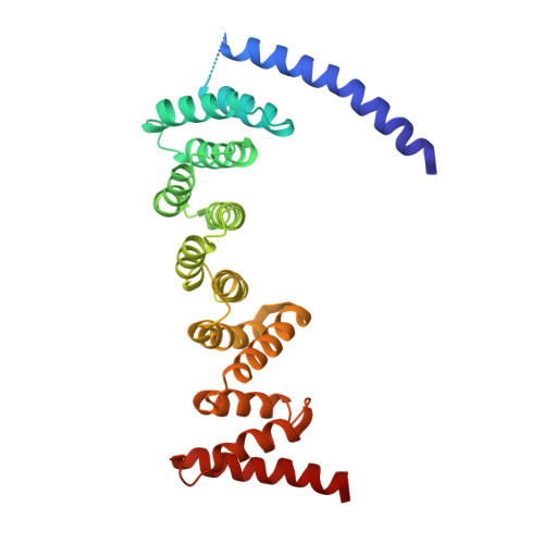
    The TRAPP complexes are nucleotide exchange factors that play essential roles in membrane traffic and autophagy. TRAPPII activates Rab11, and TRAPPIII activates Rab1, with the two complexes sharing a core of small subunits that affect nucleotide exchange but being distinguished by specific large subunits that are essential for activity in vivo. Crystal structures of core subunits have revealed the mechanism of Rab activation, but how the core and the large subunits assemble to form the complexes is unknown. We report a cryo-EM structure of the entire Drosophila TRAPPIII complex. The TRAPPIII-specific subunits TRAPPC8 and TRAPPC11 hold the catalytic core like a pair of tongs, with TRAPPC12 and TRAPPC13 positioned at the joint between them. TRAPPC2 and TRAPPC2L link the core to the two large arms, with the interfaces containing residues affected by disease-causing mutations. The TRAPPC8 arm is positioned such that it would contact Rab1 that is bound to the core, indicating how the arm could determine the specificity of the complex. A lower resolution structure of TRAPPII shows a similar architecture and suggests that the TRAPP complexes evolved from a single ur-TRAPP.


  • Organizational Affiliation

    MRC Laboratory of Molecular Biology, Cambridge, UK.


Macromolecules
Find similar proteins by:  (by identity cutoff)  |  3D Structure
Entity ID: 1
MoleculeChains Sequence LengthOrganismDetailsImage
FI18195p1307Drosophila melanogasterMutation(s): 0 
Gene Names: l(3)76BDmDmel\CG8793l(3)76BDm-RAl(3)L3809TRAPPC8CG8793Dmel_CG8793
UniProt
Find proteins for Q9VW22 (Drosophila melanogaster)
Explore Q9VW22 
Go to UniProtKB:  Q9VW22
Entity Groups  
Sequence Clusters30% Identity50% Identity70% Identity90% Identity95% Identity100% Identity
UniProt GroupQ9VW22
Sequence Annotations
Expand
  • Reference Sequence
Experimental Data & Validation

Experimental Data

  • Method: ELECTRON MICROSCOPY
  • Resolution: 4.50 Å
  • Aggregation State: PARTICLE 
  • Reconstruction Method: SINGLE PARTICLE 
EM Software:
TaskSoftware PackageVersion
RECONSTRUCTIONRELION3.1.1
MODEL REFINEMENTPHENIX

Structure Validation

View Full Validation Report


Entry History 

Deposition Data

Revision History  (Full details and data files)

  • Version 1.0: 2021-06-09
    Type: Initial release
  • Version 1.1: 2021-06-16
    Changes: Database references
  • Version 2.0: 2021-06-23
    Changes: Advisory, Atomic model, Database references, Derived calculations, Polymer sequence, Source and taxonomy, Structure summary
  • Version 2.1: 2021-07-07
    Changes: Database references