8AO9

Specific covalent inhibitor(10) of ERK2


Experimental Data Snapshot

  • Method: X-RAY DIFFRACTION
  • Resolution: 1.62 Å
  • R-Value Free: 0.232 
  • R-Value Work: 0.181 
  • R-Value Observed: 0.184 

wwPDB Validation   3D Report Full Report


Ligand Structure Quality Assessment 


This is version 1.1 of the entry. See complete history


Literature

X-ray Screening of an Electrophilic Fragment Library and Application toward the Development of a Novel ERK 1/2 Covalent Inhibitor.

St Denis, J.D.Chessari, G.Cleasby, A.Cons, B.D.Cowan, S.Dalton, S.E.East, C.Murray, C.W.O'Reilly, M.Peakman, T.Rapti, M.Stow, J.L.

(2022) J Med Chem 65: 12319-12333

  • DOI: https://doi.org/10.1021/acs.jmedchem.2c01044

  • PubMed Abstract: 

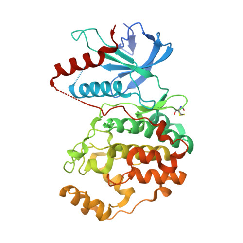
    Fragment-based drug discovery (FBDD) has become an established method for the identification of efficient starting points for drug discovery programs. In recent years, electrophilic fragment screening has garnered increased attention from both academia and industry to identify novel covalent hits for tool compound or drug development against challenging drug targets. Herein, we describe the design and characterization of an acrylamide-focused electrophilic fragment library and screening campaign against extracellular signal-regulated kinase 2 (ERK2) using high-throughput protein crystallography as the primary hit-finding technology. Several fragments were found to have covalently modified the adenosine triphosphate (ATP) binding pocket Cys166 residue. From these hits, 22 , a covalent ATP-competitive inhibitor with improved potency (ERK2 IC 50 = 7.8 μM), was developed.


  • Organizational Affiliation

    Astex Pharmaceuticals, 436 Cambridge Science Park, Cambridge CB4 0QA, United Kingdom.


Macromolecules
Find similar proteins by:  (by identity cutoff)  |  3D Structure
Entity ID: 1
MoleculeChains Sequence LengthOrganismDetailsImage
Mitogen-activated protein kinase 1368Homo sapiensMutation(s): 0 
Gene Names: MAPK1ERK2PRKM1PRKM2
EC: 2.7.11.24
UniProt & NIH Common Fund Data Resources
Find proteins for P28482 (Homo sapiens)
Explore P28482 
Go to UniProtKB:  P28482
PHAROS:  P28482
GTEx:  ENSG00000100030 
Entity Groups  
Sequence Clusters30% Identity50% Identity70% Identity90% Identity95% Identity100% Identity
UniProt GroupP28482
Sequence Annotations
Expand
  • Reference Sequence
Small Molecules
Ligands 4 Unique
IDChains Name / Formula / InChI Key2D Diagram3D Interactions
N4F (Subject of Investigation/LOI)
Query on N4F

Download Ideal Coordinates CCD File 
G [auth A]1-(6,7-dihydro-4~{H}-[1,3]thiazolo[5,4-c]pyridin-5-yl)propan-1-one
C9 H12 N2 O S
AIGPRWNLVUWLJG-UHFFFAOYSA-N
PEG
Query on PEG

Download Ideal Coordinates CCD File 
D [auth A],
E [auth A]
DI(HYDROXYETHYL)ETHER
C4 H10 O3
MTHSVFCYNBDYFN-UHFFFAOYSA-N
SO4
Query on SO4

Download Ideal Coordinates CCD File 
B [auth A]SULFATE ION
O4 S
QAOWNCQODCNURD-UHFFFAOYSA-L
EDO
Query on EDO

Download Ideal Coordinates CCD File 
C [auth A],
F [auth A]
1,2-ETHANEDIOL
C2 H6 O2
LYCAIKOWRPUZTN-UHFFFAOYSA-N
Modified Residues  1 Unique
IDChains TypeFormula2D DiagramParent
CME
Query on CME
A
L-PEPTIDE LINKINGC5 H11 N O3 S2CYS
Experimental Data & Validation

Experimental Data

  • Method: X-RAY DIFFRACTION
  • Resolution: 1.62 Å
  • R-Value Free: 0.232 
  • R-Value Work: 0.181 
  • R-Value Observed: 0.184 
  • Space Group: P 1 21 1
Unit Cell:
Length ( Å )Angle ( ˚ )
a = 48.701α = 90
b = 70.44β = 109.2
c = 60.445γ = 90
Software Package:
Software NamePurpose
BUSTERrefinement
PDB_EXTRACTdata extraction
XDSdata reduction
Aimlessdata scaling
BUSTERphasing

Structure Validation

View Full Validation Report


Ligand Structure Quality Assessment 


Entry History & Funding Information

Deposition Data

  • Released Date: 2022-09-28 
  • Deposition Author(s): Cleasby, A.

Funding OrganizationLocationGrant Number
Not fundedUnited Kingdom--

Revision History  (Full details and data files)

  • Version 1.0: 2022-09-28
    Type: Initial release
  • Version 1.1: 2022-10-05
    Changes: Database references