8C2R | pdb_00008c2r

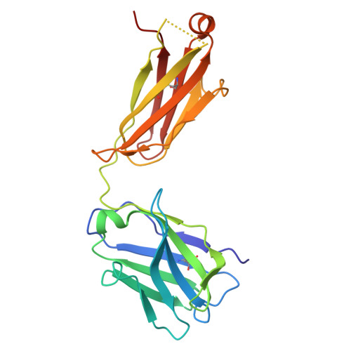
SARS-CoV2 Omicron BA.1 spike in complex with CAB-A17 antibody


Experimental Data Snapshot

  • Method: ELECTRON MICROSCOPY
  • Resolution: 2.56 Å
  • Aggregation State: PARTICLE 
  • Reconstruction Method: SINGLE PARTICLE 

Starting Model: experimental
View more details

wwPDB Validation   3D Report Full Report


This is version 1.3 of the entry. See complete history


Literature

Structural basis of broad SARS-CoV-2 cross-neutralization by affinity-matured public antibodies.

Sheward, D.J.Pushparaj, P.Das, H.Greaney, A.J.Kim, C.Kim, S.Hanke, L.Hyllner, E.Dyrdak, R.Lee, J.Dopico, X.C.Dosenovic, P.Peacock, T.P.McInerney, G.M.Albert, J.Corcoran, M.Bloom, J.D.Murrell, B.Karlsson Hedestam, G.B.Hallberg, B.M.

(2024) Cell Rep Med 5: 101577-101577

  • DOI: https://doi.org/10.1016/j.xcrm.2024.101577
  • Primary Citation of Related Structures:  
    8C0Y, 8C2R, 8V4F

  • PubMed Abstract: 

    Descendants of the severe acute respiratory syndrome coronavirus 2 (SARS-CoV-2) Omicron variant now account for almost all SARS-CoV-2 infections. The Omicron variant and its sublineages have spike glycoproteins that are highly diverged from the pandemic founder and first-generation vaccine strain, resulting in significant evasion from monoclonal antibody therapeutics and vaccines. Understanding how commonly elicited antibodies can broaden to cross-neutralize escape variants is crucial. We isolate IGHV3-53, using "public" monoclonal antibodies (mAbs) from an individual 7 months post infection with the ancestral virus and identify antibodies that exhibit potent and broad cross-neutralization, extending to the BA.1, BA.2, and BA.4/BA.5 sublineages of Omicron. Deep mutational scanning reveals these mAbs' high resistance to viral escape. Structural analysis via cryoelectron microscopy of a representative broadly neutralizing antibody, CAB-A17, in complex with the Omicron BA.1 spike highlights the structural underpinnings of this broad neutralization. By reintroducing somatic hypermutations into a germline-reverted CAB-A17, we delineate the role of affinity maturation in the development of cross-neutralization by a public class of antibodies.


  • Organizational Affiliation
    • Department of Microbiology, Tumor and Cell Biology, Karolinska Institutet, Stockholm, Sweden; Division of Medical Virology, Institute of Infectious Diseases and Molecular Medicine, University of Cape Town, Cape Town, South Africa.

Macromolecules
Find similar proteins by:  (by identity cutoff)  |  3D Structure
Entity ID: 1
MoleculeChains Sequence LengthOrganismDetailsImage
Spike glycoprotein
A, B, C
1,122Severe acute respiratory syndrome coronavirus 2Mutation(s): 33 
Gene Names: S2
UniProt
Find proteins for P0DTC2 (Severe acute respiratory syndrome coronavirus 2)
Explore P0DTC2 
Go to UniProtKB:  P0DTC2
Entity Groups  
Sequence Clusters30% Identity50% Identity70% Identity90% Identity95% Identity100% Identity
UniProt GroupP0DTC2
Glycosylation
Glycosylation Sites: 6Go to GlyGen: P0DTC2-1
Sequence Annotations
Expand
  • Reference Sequence
Find similar proteins by:  (by identity cutoff)  |  3D Structure
Entity ID: 2
MoleculeChains Sequence LengthOrganismDetailsImage
CAB-A17 antibody (LC)D [auth E],
F [auth L]
214Homo sapiensMutation(s): 0 
Entity Groups  
Sequence Clusters30% Identity50% Identity70% Identity90% Identity95% Identity100% Identity
Sequence Annotations
Expand
  • Reference Sequence
Find similar proteins by:  (by identity cutoff)  |  3D Structure
Entity ID: 3
MoleculeChains Sequence LengthOrganismDetailsImage
CAB-A17 antibody (HC)E [auth F],
G [auth R]
213Homo sapiensMutation(s): 0 
Entity Groups  
Sequence Clusters30% Identity50% Identity70% Identity90% Identity95% Identity100% Identity
Sequence Annotations
Expand
  • Reference Sequence
Small Molecules
Ligands 1 Unique
IDChains Name / Formula / InChI Key2D Diagram3D Interactions
NAG
Query on NAG

Download Ideal Coordinates CCD File 
AA [auth A]
BA [auth A]
CA [auth A]
DA [auth A]
EA [auth B]
AA [auth A],
BA [auth A],
CA [auth A],
DA [auth A],
EA [auth B],
FA [auth B],
GA [auth B],
HA [auth B],
IA [auth B],
JA [auth B],
KA [auth B],
LA [auth B],
MA [auth C],
NA [auth C],
OA [auth C],
PA [auth C],
QA [auth C],
RA [auth C],
W [auth A],
X [auth A],
Y [auth A],
Z [auth A]
2-acetamido-2-deoxy-beta-D-glucopyranose
C8 H15 N O6
OVRNDRQMDRJTHS-FMDGEEDCSA-N
Experimental Data & Validation

Experimental Data

  • Method: ELECTRON MICROSCOPY
  • Resolution: 2.56 Å
  • Aggregation State: PARTICLE 
  • Reconstruction Method: SINGLE PARTICLE 
EM Software:
TaskSoftware PackageVersion
MODEL REFINEMENTPHENIX1.19
RECONSTRUCTIONcryoSPARC3.3.1

Structure Validation

View Full Validation Report


Entry History & Funding Information

Deposition Data


Funding OrganizationLocationGrant Number
Not funded--

Revision History  (Full details and data files)

  • Version 1.0: 2024-05-22
    Type: Initial release
  • Version 1.1: 2024-09-25
    Changes: Data collection, Source and taxonomy
  • Version 1.2: 2024-11-06
    Changes: Data collection, Structure summary
  • Version 1.3: 2024-12-04
    Changes: Data collection, Database references