8DEI | pdb_00008dei

Structure of the Cac1 KER domain


Experimental Data Snapshot

  • Method: X-RAY DIFFRACTION
  • Resolution: 2.81 Å
  • R-Value Free: 
    0.282 (Depositor), 0.280 (DCC) 
  • R-Value Work: 
    0.233 (Depositor), 0.230 (DCC) 
  • R-Value Observed: 
    0.238 (Depositor) 

Starting Model: experimental
View more details

wwPDB Validation   3D Report Full Report


Ligand Structure Quality Assessment 


This is version 1.3 of the entry. See complete history


Literature

A novel single alpha-helix DNA-binding domain in CAF-1 promotes gene silencing and DNA damage survival through tetrasome-length DNA selectivity and spacer function.

Rosas, R.Aguilar, R.R.Arslanovic, N.Seck, A.Smith, D.J.Tyler, J.K.Churchill, M.E.A.

(2023) Elife 12

  • DOI: https://doi.org/10.7554/eLife.83538
  • Primary Citation of Related Structures:  
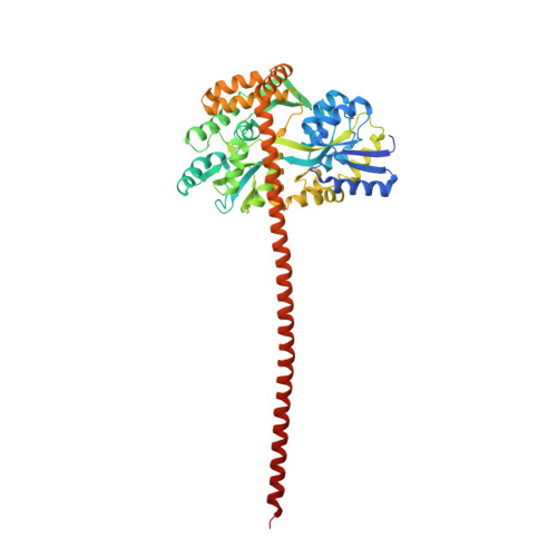
    8DEI

  • PubMed Abstract: 

    The histone chaperone chromatin assembly factor 1 (CAF-1) deposits two nascent histone H3/H4 dimers onto newly replicated DNA forming the central core of the nucleosome known as the tetrasome. How CAF-1 ensures there is sufficient space for the assembly of tetrasomes remains unknown. Structural and biophysical characterization of the lysine/glutamic acid/arginine-rich (KER) region of CAF-1 revealed a 128-Å single alpha-helix (SAH) motif with unprecedented DNA-binding properties. Distinct KER sequence features and length of the SAH drive the selectivity of CAF-1 for tetrasome-length DNA and facilitate function in budding yeast. In vivo, the KER cooperates with the DNA-binding winged helix domain in CAF-1 to overcome DNA damage sensitivity and maintain silencing of gene expression. We propose that the KER SAH links functional domains within CAF-1 with structural precision, acting as a DNA-binding spacer element during chromatin assembly.


  • Organizational Affiliation
    • Program in Structural Biology and Biochemistry, University of Colorado Anschutz Medical Campus, Aurora, United States.

Macromolecules
Find similar proteins by:  (by identity cutoff)  |  3D Structure
Entity ID: 1
MoleculeChains Sequence LengthOrganismDetailsImage
Maltodextrin-binding protein,Chromatin assembly factor 1 subunit p90 fusion
A, B, C, D
490Escherichia coliSaccharomyces cerevisiae
This entity is chimeric
Mutation(s): 0 
Gene Names: malEmalE_1malE_2RLF2CAC1YPR018WYP9531.12
UniProt
Find proteins for P0AEX9 (Escherichia coli (strain K12))
Explore P0AEX9 
Go to UniProtKB:  P0AEX9
Find proteins for Q12495 (Saccharomyces cerevisiae (strain ATCC 204508 / S288c))
Explore Q12495 
Go to UniProtKB:  Q12495
Entity Groups  
Sequence Clusters30% Identity50% Identity70% Identity90% Identity95% Identity100% Identity
UniProt GroupsQ12495P0AEX9
Sequence Annotations
Expand
  • Reference Sequence
Small Molecules
Ligands 3 Unique
IDChains Name / Formula / InChI Key2D Diagram3D Interactions
PEG (Subject of Investigation/LOI)
Query on PEG

Download Ideal Coordinates CCD File 
E [auth B],
F [auth B],
I [auth C],
J [auth C],
K [auth D]
DI(HYDROXYETHYL)ETHER
C4 H10 O3
MTHSVFCYNBDYFN-UHFFFAOYSA-N
PO4
Query on PO4

Download Ideal Coordinates CCD File 
G [auth C]PHOSPHATE ION
O4 P
NBIIXXVUZAFLBC-UHFFFAOYSA-K
GOL
Query on GOL

Download Ideal Coordinates CCD File 
H [auth C]GLYCEROL
C3 H8 O3
PEDCQBHIVMGVHV-UHFFFAOYSA-N
Experimental Data & Validation

Experimental Data

  • Method: X-RAY DIFFRACTION
  • Resolution: 2.81 Å
  • R-Value Free:  0.282 (Depositor), 0.280 (DCC) 
  • R-Value Work:  0.233 (Depositor), 0.230 (DCC) 
  • R-Value Observed: 0.238 (Depositor) 
Space Group: P 1 21 1
Unit Cell:
Length ( Å )Angle ( ˚ )
a = 53.586α = 90
b = 165.249β = 96.475
c = 116.149γ = 90
Software Package:
Software NamePurpose
Cootmodel building
PHENIXrefinement
CrysalisProdata reduction
Aimlessdata scaling
PHASERphasing

Structure Validation

View Full Validation Report


Ligand Structure Quality Assessment 


Entry History & Funding Information

Deposition Data


Funding OrganizationLocationGrant Number
National Institutes of Health/National Institute of General Medical Sciences (NIH/NIGMS)United StatesR01 GM135604

Revision History  (Full details and data files)

  • Version 1.0: 2023-07-05
    Type: Initial release
  • Version 1.1: 2023-10-04
    Changes: Data collection, Database references, Structure summary
  • Version 1.2: 2023-10-25
    Changes: Refinement description
  • Version 1.3: 2024-02-14
    Changes: Database references