Tuning the Metabolic Stability of Visual Cycle Modulators through Modification of an RPE65 Recognition Motif.
Bassetto, M., Zaluski, J., Li, B., Zhang, J., Badiee, M., Kiser, P.D., Tochtrop, G.P.(2023) J Med Chem 66: 8140-8158
- PubMed: 37279401
- DOI: https://doi.org/10.1021/acs.jmedchem.3c00461
- Primary Citation of Related Structures:
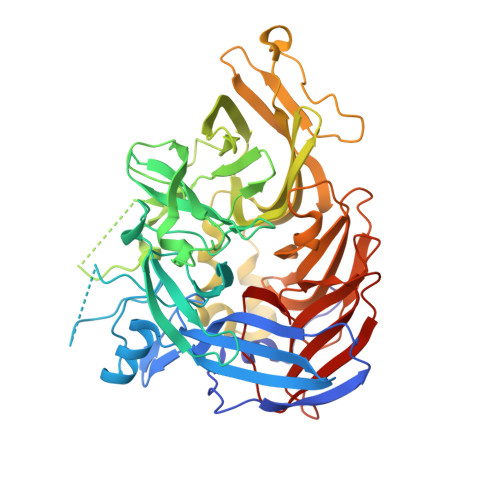
8DOC - PubMed Abstract:
In the eye, the isomerization of all- trans -retinal to 11- cis -retinal is accomplished by a metabolic pathway termed the visual cycle that is critical for vision. RPE65 is the essential trans - cis isomerase of this pathway. Emixustat, a retinoid-mimetic RPE65 inhibitor, was developed as a therapeutic visual cycle modulator and used for the treatment of retinopathies. However, pharmacokinetic liabilities limit its further development including: (1) metabolic deamination of the γ-amino-α-aryl alcohol, which mediates targeted RPE65 inhibition, and (2) unwanted long-lasting RPE65 inhibition. We sought to address these issues by more broadly defining the structure-activity relationships of the RPE65 recognition motif via the synthesis of a family of novel derivatives, which were tested in vitro and in vivo for RPE65 inhibition. We identified a potent secondary amine derivative with resistance to deamination and preserved RPE65 inhibitory activity. Our data provide insights into activity-preserving modifications of the emixustat molecule that can be employed to tune its pharmacological properties.
- Department of Physiology and Biophysics, School of Medicine, University of California - Irvine, Irvine, California 92697, United States.
Organizational Affiliation: