8HMA | pdb_00008hma

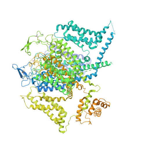
Cryo-EM structure of human high-voltage activated L-type calcium channel CaV1.2 in complex with tetrandrine (TET)


Experimental Data Snapshot

  • Method: ELECTRON MICROSCOPY
  • Resolution: 3.40 Å
  • Aggregation State: PARTICLE 
  • Reconstruction Method: SINGLE PARTICLE 

wwPDB Validation   3D Report Full Report


This is version 1.2 of the entry. See complete history


Literature

Structural bases of inhibitory mechanism of Ca V 1.2 channel inhibitors.

Wei, Y.Yu, Z.Wang, L.Li, X.Li, N.Bai, Q.Wang, Y.Li, R.Meng, Y.Xu, H.Wang, X.Dong, Y.Huang, Z.Zhang, X.C.Zhao, Y.

(2024) Nat Commun 15: 2772-2772

  • DOI: https://doi.org/10.1038/s41467-024-47116-8
  • Primary Citation of Related Structures:  
    8HLP, 8HMA, 8HMB

  • PubMed Abstract: 

    The voltage-gated calcium channel Ca V 1.2 is essential for cardiac and vessel smooth muscle contractility and brain function. Accumulating evidence demonstrates that malfunctions of Ca V 1.2 are involved in brain and heart diseases. Pharmacological inhibition of Ca V 1.2 is therefore of therapeutic value. Here, we report cryo-EM structures of Ca V 1.2 in the absence or presence of the antirheumatic drug tetrandrine or antihypertensive drug benidipine. Tetrandrine acts as a pore blocker in a pocket composed of S6 II , S6 III , and S6 IV helices and forms extensive hydrophobic interactions with Ca V 1.2. Our structure elucidates that benidipine is located in the D III -D IV fenestration site. Its hydrophobic sidechain, phenylpiperidine, is positioned at the exterior of the pore domain and cradled within a hydrophobic pocket formed by S5 DIII , S6 DIII , and S6 DIV helices, providing additional interactions to exert inhibitory effects on both L-type and T-type voltage gated calcium channels. These findings provide the structural foundation for the rational design and optimization of therapeutic inhibitors of voltage-gated calcium channels.


  • Organizational Affiliation
    • Key Laboratory of Biomacromolecules (CAS), National Laboratory of Biomacromolecules, CAS Center for Excellence in Biomacromolecules, Institute of Biophysics, Chinese Academy of Sciences, Beijing, 100101, China.

Macromolecules
Find similar proteins by:  (by identity cutoff)  |  3D Structure
Entity ID: 1
MoleculeChains Sequence LengthOrganismDetailsImage
Voltage-dependent L-type calcium channel subunit alphaA [auth E]2,173Homo sapiensMutation(s): 0 
Gene Names: CACNA1C
Membrane Entity: Yes 
UniProt & NIH Common Fund Data Resources
Find proteins for Q13936 (Homo sapiens)
Explore Q13936 
Go to UniProtKB:  Q13936
PHAROS:  Q13936
GTEx:  ENSG00000151067 
Entity Groups  
Sequence Clusters30% Identity50% Identity70% Identity90% Identity95% Identity100% Identity
UniProt GroupQ13936
Sequence Annotations
Expand
  • Reference Sequence
Find similar proteins by:  (by identity cutoff)  |  3D Structure
Entity ID: 2
MoleculeChains Sequence LengthOrganismDetailsImage
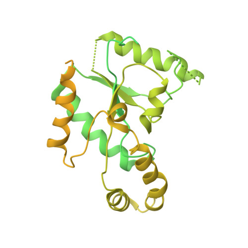
Isoform 2c of Voltage-dependent L-type calcium channel subunit beta-2B [auth H]632Homo sapiensMutation(s): 0 
Gene Names: CACNB2CACNLB2MYSB
Membrane Entity: Yes 
UniProt & NIH Common Fund Data Resources
Find proteins for Q08289 (Homo sapiens)
Explore Q08289 
Go to UniProtKB:  Q08289
PHAROS:  Q08289
GTEx:  ENSG00000165995 
Entity Groups  
Sequence Clusters30% Identity50% Identity70% Identity90% Identity95% Identity100% Identity
UniProt GroupQ08289
Sequence Annotations
Expand
  • Reference Sequence
Find similar proteins by:  (by identity cutoff)  |  3D Structure
Entity ID: 3
MoleculeChains Sequence LengthOrganismDetailsImage
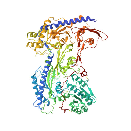
Voltage-dependent calcium channel subunit alpha-2/delta-1C [auth F]1,071Homo sapiensMutation(s): 0 
Gene Names: CACNA2D1CACNL2ACCHL2AMHS3
Membrane Entity: Yes 
UniProt & NIH Common Fund Data Resources
Find proteins for P54289 (Homo sapiens)
Explore P54289 
Go to UniProtKB:  P54289
PHAROS:  P54289
GTEx:  ENSG00000153956 
Entity Groups  
Sequence Clusters30% Identity50% Identity70% Identity90% Identity95% Identity100% Identity
UniProt GroupP54289
Glycosylation
Glycosylation Sites: 9Go to GlyGen: P54289-1
Sequence Annotations
Expand
  • Reference Sequence
Small Molecules
Ligands 6 Unique
IDChains Name / Formula / InChI Key2D Diagram3D Interactions
3PE
Query on 3PE

Download Ideal Coordinates CCD File 
E,
F [auth E],
H [auth E]
1,2-Distearoyl-sn-glycerophosphoethanolamine
C41 H82 N O8 P
LVNGJLRDBYCPGB-LDLOPFEMSA-N
A1AC9 (Subject of Investigation/LOI)
Query on A1AC9

Download Ideal Coordinates CCD File 
G [auth E]6,6',7,12-tetramethoxy-2,2'-dimethyl-1beta-3,4-didehydroberbaman
C38 H40 N2 O6
OUHWUXGBWHCKDH-UFXYQILXSA-N
R16
Query on R16

Download Ideal Coordinates CCD File 
D [auth E]HEXADECANE
C16 H34
DCAYPVUWAIABOU-UHFFFAOYSA-N
NAG
Query on NAG

Download Ideal Coordinates CCD File 
K [auth F]
L [auth F]
M [auth F]
N [auth F]
P [auth F]
K [auth F],
L [auth F],
M [auth F],
N [auth F],
P [auth F],
Q [auth F],
R [auth F],
S [auth F],
T [auth F],
U [auth F],
V [auth F]
2-acetamido-2-deoxy-beta-D-glucopyranose
C8 H15 N O6
OVRNDRQMDRJTHS-FMDGEEDCSA-N
BMA
Query on BMA

Download Ideal Coordinates CCD File 
O [auth F]beta-D-mannopyranose
C6 H12 O6
WQZGKKKJIJFFOK-RWOPYEJCSA-N
CA
Query on CA

Download Ideal Coordinates CCD File 
I [auth E],
J [auth E]
CALCIUM ION
Ca
BHPQYMZQTOCNFJ-UHFFFAOYSA-N
Experimental Data & Validation

Experimental Data

  • Method: ELECTRON MICROSCOPY
  • Resolution: 3.40 Å
  • Aggregation State: PARTICLE 
  • Reconstruction Method: SINGLE PARTICLE 
EM Software:
TaskSoftware PackageVersion
MODEL REFINEMENTPHENIX

Structure Validation

View Full Validation Report


Entry History & Funding Information

Deposition Data


Funding OrganizationLocationGrant Number
Chinese Academy of SciencesChina--

Revision History  (Full details and data files)

  • Version 1.0: 2024-04-24
    Type: Initial release
  • Version 1.1: 2024-11-06
    Changes: Data collection, Structure summary
  • Version 1.2: 2025-06-04
    Changes: Data collection