8PKB | pdb_00008pkb

Staphylococcus aureus endonuclease IV with bound phosphate


Experimental Data Snapshot

  • Method: X-RAY DIFFRACTION
  • Resolution: 1.90 Å
  • R-Value Free: 
    0.211 (Depositor), 0.210 (DCC) 
  • R-Value Work: 
    0.188 (Depositor), 0.190 (DCC) 
  • R-Value Observed: 
    0.189 (Depositor) 

Starting Model: experimental
View more details

wwPDB Validation   3D Report Full Report


Ligand Structure Quality Assessment 


This is version 1.2 of the entry. See complete history


Literature

Octahedral Iron in Catalytic Sites of Endonuclease IV from Staphylococcus aureus and Escherichia coli .

Kirillov, S.Isupov, M.Paterson, N.G.Wiener, R.Abeldenov, S.Saper, M.A.Rouvinski, A.

(2025) Biochemistry 64: 67-82

  • DOI: https://doi.org/10.1021/acs.biochem.4c00447
  • Primary Citation of Related Structures:  
    8AXY, 8EDD, 8PKB, 8RLY

  • PubMed Abstract: 

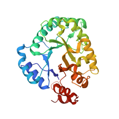
    During Staphylococcus aureus infections, reactive oxygen species cause DNA damage, including nucleotide base modification. After removal of the defective base, excision repair requires an endonuclease IV (Nfo), which hydrolyzes the phosphodiester bond 5' to the abasic nucleotide. This class of enzymes, typified by the enzyme from Escherichia coli , contains a catalytic site with three metal ions, previously reported to be all Zn 2+ . The 1.05 Å structure of Nfo from the Gram-positive organism S. aureus ( Sa Nfo) revealed two inner Fe 2+ ions and one Zn 2+ as confirmed by dispersive anomalous difference maps. Sa Nfo has a previously undescribed water molecule liganded to Fe 1 forming an octahedral coordination geometry and hydrogen bonded to Tyr33, an active site residue conserved in many Gram-positive bacteria, but which is Phe in Gram-negative species that coordinate Zn 2+ at the corresponding site. The 1.9 Å structure of E. coli Nfo ( Ec Nfo), purified without added metals, revealed that metal 2 is Fe 2+ and not Zn 2+ . Octahedral coordination for the sites occupied by Fe 2+ suggests a stereoselective mechanism for differentiating between Fe 2+ and Zn 2+ in this enzyme class. Kinetics and an inhibitor competition assay of Sa Nfo reveal product inhibition (or slow product release), especially at low ionic strength, caused in part by a Lys-rich DNA binding loop present in Sa Nfo and Gram-positive species but not in Ec Nfo. Biological significance of the slow product release is discussed. Catalytic activity in vitro is optimal at 300 mM NaCl, which is consistent with the halotolerant phenotype of S. aureus .


  • Organizational Affiliation
    • Department of General Biology and Genomics, L. N. Gumilyov Eurasian National University, Astana 010008, Kazakhstan.

Macromolecules
Find similar proteins by:  (by identity cutoff)  |  3D Structure
Entity ID: 1
MoleculeChains Sequence LengthOrganismDetailsImage
Probable endonuclease 4296Staphylococcus aureusMutation(s): 0 
Gene Names: nfoSAR1634
EC: 3.1.21.2
UniProt
Find proteins for Q6GGE2 (Staphylococcus aureus (strain MRSA252))
Explore Q6GGE2 
Go to UniProtKB:  Q6GGE2
Entity Groups  
Sequence Clusters30% Identity50% Identity70% Identity90% Identity95% Identity100% Identity
UniProt GroupQ6GGE2
Sequence Annotations
Expand
  • Reference Sequence
Small Molecules
Ligands 4 Unique
IDChains Name / Formula / InChI Key2D Diagram3D Interactions
PO4 (Subject of Investigation/LOI)
Query on PO4

Download Ideal Coordinates CCD File 
E [auth A]PHOSPHATE ION
O4 P
NBIIXXVUZAFLBC-UHFFFAOYSA-K
ZN (Subject of Investigation/LOI)
Query on ZN

Download Ideal Coordinates CCD File 
D [auth A]ZINC ION
Zn
PTFCDOFLOPIGGS-UHFFFAOYSA-N
FE (Subject of Investigation/LOI)
Query on FE

Download Ideal Coordinates CCD File 
B [auth A],
C [auth A]
FE (III) ION
Fe
VTLYFUHAOXGGBS-UHFFFAOYSA-N
CL
Query on CL

Download Ideal Coordinates CCD File 
F [auth A],
G [auth A]
CHLORIDE ION
Cl
VEXZGXHMUGYJMC-UHFFFAOYSA-M
Experimental Data & Validation

Experimental Data

  • Method: X-RAY DIFFRACTION
  • Resolution: 1.90 Å
  • R-Value Free:  0.211 (Depositor), 0.210 (DCC) 
  • R-Value Work:  0.188 (Depositor), 0.190 (DCC) 
  • R-Value Observed: 0.189 (Depositor) 
Space Group: P 21 21 21
Unit Cell:
Length ( Å )Angle ( ˚ )
a = 38.548α = 90
b = 84.04β = 90
c = 103.344γ = 90
Software Package:
Software NamePurpose
PHENIXrefinement
XDSdata reduction
Aimlessdata scaling
PHASERphasing

Structure Validation

View Full Validation Report


Ligand Structure Quality Assessment 


Entry History & Funding Information

Deposition Data


Funding OrganizationLocationGrant Number
Other government--

Revision History  (Full details and data files)

  • Version 1.0: 2024-07-10
    Type: Initial release
  • Version 1.1: 2024-12-25
    Changes: Database references, Structure summary
  • Version 1.2: 2025-01-15
    Changes: Database references