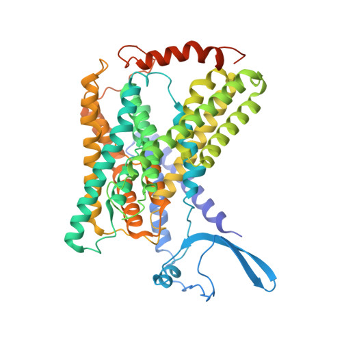
PIP 2 -mediated oligomerization of the endosomal sodium/proton exchanger NHE9.
Kokane, S., Gulati, A., Meier, P.F., Matsuoka, R., Pipatpolkai, T., Albano, G., Ho, T.M., Delemotte, L., Fuster, D., Drew, D.(2025) Nat Commun 16: 3055-3055
- PubMed: 40155618
- DOI: https://doi.org/10.1038/s41467-025-58247-x
- Primary Citation of Related Structures:
8PS0, 8PVR, 8PXB - PubMed Abstract:
The strict exchange of Na + for H + ions across cell membranes is a reaction carried out in almost every cell. Na + /H + exchangers that perform this task are physiological homodimers, and whilst the ion transporting domain is highly conserved, their dimerization differs. The Na + /H + exchanger NhaA from Escherichia coli has a weak dimerization interface mediated by a β-hairpin domain and with dimer retention dependent on cardiolipin. Similarly, organellar Na + /H + exchangers NHE6, NHE7 and NHE9 also contain β-hairpin domains and recent analysis of Equus caballus NHE9 indicated PIP 2 lipids could bind at the dimer interface. However, structural validation of the predicted lipid-mediated oligomerization has been lacking. Here, we report cryo-EM structures of E. coli NhaA and E. caballus NHE9 in complex with cardiolipin and phosphatidylinositol-3,5-bisphosphate PI(3,5)P 2 lipids binding at their respective dimer interfaces. We further show how the endosomal specific PI(3,5)P 2 lipid stabilizes the NHE9 homodimer and enhances transport activity. Indeed, we show that NHE9 is active in endosomes, but not at the plasma membrane where the PI(3,5)P 2 lipid is absent. Thus, specific lipids can regulate Na + /H + exchange activity by stabilizing dimerization in response to either cell specific cues or upon trafficking to their correct membrane location.
- Department of Biochemistry and Biophysics, Science for Life laboratory, Stockholm University, Stockholm, Sweden.
Organizational Affiliation: