8RJQ | pdb_00008rjq

Serial femtosecond X-ray structure of a fluorescence optimized bathy phytochrome PAiRFP2 derived from wild-type Agp2 in I3 intermediate state.


Experimental Data Snapshot

  • Method: X-RAY DIFFRACTION
  • Resolution: 2.40 Å
  • R-Value Free: 
    0.221 (Depositor), 0.220 (DCC) 
  • R-Value Work: 
    0.203 (Depositor), 0.207 (DCC) 
  • R-Value Observed: 
    0.204 (Depositor) 

Starting Model: experimental
View more details

wwPDB Validation   3D Report Full Report


Ligand Structure Quality Assessment 


This is version 1.1 of the entry. See complete history


Literature

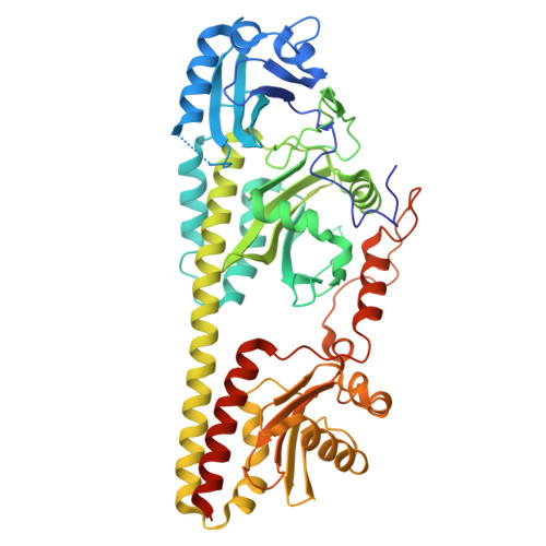
Serial-femtosecond crystallography reveals how a phytochrome variant couples chromophore and protein structural changes.

Sauthof, L.Szczepek, M.Schmidt, A.Bhowmick, A.Dasgupta, M.Mackintosh, M.J.Gul, S.Fuller, F.D.Chatterjee, R.Young, I.D.Michael, N.Heyder, N.A.Bauer, B.Koch, A.Bogacz, I.Kim, I.S.Simon, P.S.Butryn, A.Aller, P.Chukhutsina, V.U.Baxter, J.M.Hutchison, C.D.M.Liebschner, D.Poon, B.Sauter, N.K.Miller, M.D.Alonso-Mori, R.Hunter, M.S.Batyuk, A.Owada, S.Tono, K.Tanaka, R.van Thor, J.J.Krauss, N.Lamparter, T.Brewster, A.S.Schapiro, I.Orville, A.M.Yachandra, V.K.Yano, J.Hildebrandt, P.Kern, J.F.Scheerer, P.

(2025) Sci Adv 11: eadp2665-eadp2665

  • DOI: https://doi.org/10.1126/sciadv.adp2665
  • Primary Citation of Related Structures:  
    8RJM, 8RJN, 8RJO, 8RJP, 8RJQ, 8RJR, 8RJS, 8RJT, 8RJU

  • PubMed Abstract: 

    The photoreaction and commensurate structural changes of a chromophore within biological photoreceptors elicit conformational transitions of the protein promoting the switch between deactivated and activated states. We investigated how this coupling is achieved in a bacterial phytochrome variant, Agp2-PAiRFP2. Contrary to classical protein crystallography, which only allows probing (cryo-trapped) stable states, we have used time-resolved serial femtosecond x-ray crystallography (tr-SFX) and pump-probe techniques with various illumination and delay times with respect to photoexcitation of the parent Pfr state. Thus, structural data for seven time frames were sorted into groups of molecular events along the reaction coordinate. They range from chromophore isomerization to the formation of Meta-F, the intermediate that precedes the functional relevant secondary structure transition of the tongue. Structural data for the early events were used to calculate the photoisomerization pathway to complement the experimental data. Late events allow identifying the molecular switch that is linked to the intramolecular proton transfer as a prerequisite for the following structural transitions.


  • Organizational Affiliation

    Charité-Universitätsmedizin Berlin, corporate member of Freie Universität Berlin and Humboldt-Universität zu Berlin, Institute of Medical Physics and Biophysics, Group Structural Biology of Cellular Signaling, Charitéplatz 1, D-10117, Berlin, Germany.


Macromolecules
Find similar proteins by:  (by identity cutoff)  |  3D Structure
Entity ID: 1
MoleculeChains Sequence LengthOrganismDetailsImage
histidine kinase
A, B
515Agrobacterium fabrum str. C58Mutation(s): 24 
Gene Names: Atu2165
EC: 2.7.13.3
UniProt
Find proteins for A9CI81 (Agrobacterium fabrum (strain C58 / ATCC 33970))
Explore A9CI81 
Go to UniProtKB:  A9CI81
Entity Groups  
Sequence Clusters30% Identity50% Identity70% Identity90% Identity95% Identity100% Identity
UniProt GroupA9CI81
Sequence Annotations
Expand
  • Reference Sequence
Small Molecules
Ligands 4 Unique
IDChains Name / Formula / InChI Key2D Diagram3D Interactions
EL5 (Subject of Investigation/LOI)
Query on EL5

Download Ideal Coordinates CCD File 
C [auth A],
J [auth B]
3-[(2Z)-2-({3-(2-carboxyethyl)-5-[(E)-(4-ethenyl-3-methyl-5-oxo-1,5-dihydro-2H-pyrrol-2-ylidene)methyl]-4-methyl-1H-pyrrol-2-yl}methylidene)-5-{(Z)-[(3E,4S)-3-ethylidene-4-methyl-5-oxopyrrolidin-2-ylidene]methyl}-4-methyl-2H-pyrrol-3-yl]propanoic acid
C33 H36 N4 O6
SNHIGJASYQUMKZ-IDFYGOSVSA-N
PEG
Query on PEG

Download Ideal Coordinates CCD File 
I [auth A],
P [auth B]
DI(HYDROXYETHYL)ETHER
C4 H10 O3
MTHSVFCYNBDYFN-UHFFFAOYSA-N
SO4
Query on SO4

Download Ideal Coordinates CCD File 
D [auth A]
E [auth A]
F [auth A]
K [auth B]
L [auth B]
D [auth A],
E [auth A],
F [auth A],
K [auth B],
L [auth B],
M [auth B],
N [auth B]
SULFATE ION
O4 S
QAOWNCQODCNURD-UHFFFAOYSA-L
GOL
Query on GOL

Download Ideal Coordinates CCD File 
G [auth A],
H [auth A],
O [auth B]
GLYCEROL
C3 H8 O3
PEDCQBHIVMGVHV-UHFFFAOYSA-N
Experimental Data & Validation

Experimental Data

  • Method: X-RAY DIFFRACTION
  • Resolution: 2.40 Å
  • R-Value Free:  0.221 (Depositor), 0.220 (DCC) 
  • R-Value Work:  0.203 (Depositor), 0.207 (DCC) 
  • R-Value Observed: 0.204 (Depositor) 
Space Group: P 63 2 2
Unit Cell:
Length ( Å )Angle ( ˚ )
a = 184.641α = 90
b = 184.641β = 90
c = 182.171γ = 120
Software Package:
Software NamePurpose
REFMACrefinement
DIALSdata scaling
DIALSdata reduction
PRIMEdata scaling
PHASERphasing
PHENIXrefinement
Cootmodel building

Structure Validation

View Full Validation Report


Ligand Structure Quality Assessment 


Entry History & Funding Information

Deposition Data


Funding OrganizationLocationGrant Number
German Research Foundation (DFG)GermanyCRC1078
German Research Foundation (DFG)GermanyCRC1423
German Research Foundation (DFG)GermanyEXC 311 2008/1
Boehringer Ingelheim Fonds (BIF)GermanyBIF Travel Grant
Israel Science FoundationIsrael3131/20
Department of Energy (DOE, United States)United StatesDE-AC02-76SF00515, DE-AC02-05CH11231
National Institutes of Health/Office of the DirectorUnited StatesGM05530, GM110501, GM126289, GM117126, S10 OD023453
Wellcome TrustUnited Kingdom210734/Z/18/Z
Royal SocietyUnited KingdomRSWF- R2 - 182017

Revision History  (Full details and data files)

  • Version 1.0: 2025-05-14
    Type: Initial release
  • Version 1.1: 2025-06-11
    Changes: Database references