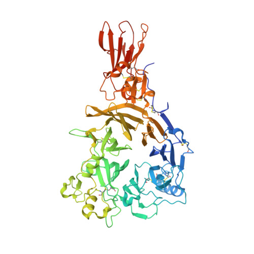
8RMJ

Drosophila Semaphorin 2b in complex with glycosaminoglycan mimic SOS


Experimental Data Snapshot

  • Method: X-RAY DIFFRACTION
  • Resolution: 2.79 Å
  • R-Value Free: 0.274 
  • R-Value Work: 0.246 
  • R-Value Observed: 0.248 

Starting Model: experimental
View more details

wwPDB Validation   3D Report Full Report


Ligand Structure Quality Assessment 


This is version 1.0 of the entry. See complete history


Literature

Molecular mechanisms of proteoglycan-mediated semaphorin signaling in axon guidance.

Nourisanami, F.Sobol, M.Li, Z.Horvath, M.Kowalska, K.Kumar, A.Vlasak, J.Koupilova, N.Luginbuhl, D.J.Luo, L.Rozbesky, D.

(2024) Proc Natl Acad Sci U S A 121: e2402755121-e2402755121

  • DOI: https://doi.org/10.1073/pnas.2402755121
  • Primary Citation of Related Structures:  
    8RMJ

  • PubMed Abstract: 

    The precise assembly of a functional nervous system relies on axon guidance cues. Beyond engaging their cognate receptors and initiating signaling cascades that modulate cytoskeletal dynamics, guidance cues also bind components of the extracellular matrix, notably proteoglycans, yet the role and mechanisms of these interactions remain poorly understood. We found that Drosophila secreted semaphorins bind specifically to glycosaminoglycan (GAG) chains of proteoglycans, showing a preference based on the degree of sulfation. Structural analysis of Sema2b unveiled multiple GAG-binding sites positioned outside canonical plexin-binding site, with the highest affinity binding site located at the C-terminal tail, characterized by a lysine-rich helical arrangement that appears to be conserved across secreted semaphorins. In vivo studies revealed a crucial role of the Sema2b C-terminal tail in specifying the trajectory of olfactory receptor neurons. We propose that secreted semaphorins tether to the cell surface through interactions with GAG chains of proteoglycans, facilitating their presentation to cognate receptors on passing axons.


  • Organizational Affiliation

    Department of Cell Biology, Faculty of Science, Charles University, Prague 128 43, Czechia.


Macromolecules
Find similar proteins by:  (by identity cutoff)  |  3D Structure
Entity ID: 1
MoleculeChains Sequence LengthOrganismDetailsImage
FI18622p1657Drosophila melanogasterMutation(s): 0 
Gene Names: Sema2bCG30322CG33960-RACG4383Dmel\CG33960Sema-2bsema2bSemaIIBCG33960Dmel_CG33960
UniProt
Find proteins for A0A0B4KG38 (Drosophila melanogaster)
Explore A0A0B4KG38 
Go to UniProtKB:  A0A0B4KG38
Entity Groups  
Sequence Clusters30% Identity50% Identity70% Identity90% Identity95% Identity100% Identity
UniProt GroupA0A0B4KG38
Glycosylation
Glycosylation Sites: 7Go to GlyGen: A0A0B4KG38-1
Sequence Annotations
Expand
  • Reference Sequence
Oligosaccharides

Help

Entity ID: 2
MoleculeChains Length2D Diagram Glycosylation3D Interactions
2-acetamido-2-deoxy-beta-D-glucopyranose-(1-4)-2-acetamido-2-deoxy-beta-D-glucopyranose
B
2N-Glycosylation
Glycosylation Resources
GlyTouCan:  G42666HT
GlyCosmos:  G42666HT
GlyGen:  G42666HT
Entity ID: 3
MoleculeChains Length2D Diagram Glycosylation3D Interactions
alpha-D-mannopyranose-(1-3)-[alpha-D-mannopyranose-(1-6)]alpha-D-mannopyranose-(1-6)-[alpha-D-mannopyranose-(1-3)]beta-D-mannopyranose-(1-4)-2-acetamido-2-deoxy-beta-D-glucopyranose-(1-4)-2-acetamido-2-deoxy-beta-D-glucopyranose
C
7N-Glycosylation
Glycosylation Resources
GlyTouCan:  G55220VL
GlyCosmos:  G55220VL
GlyGen:  G55220VL
Entity ID: 4
MoleculeChains Length2D Diagram Glycosylation3D Interactions
alpha-D-mannopyranose-(1-2)-alpha-D-mannopyranose-(1-6)-[alpha-D-mannopyranose-(1-3)]alpha-D-mannopyranose-(1-6)-[alpha-D-mannopyranose-(1-2)-alpha-D-mannopyranose-(1-3)]beta-D-mannopyranose-(1-4)-2-acetamido-2-deoxy-beta-D-glucopyranose-(1-4)-2-acetamido-2-deoxy-beta-D-glucopyranose
D
9N-Glycosylation
Glycosylation Resources
GlyTouCan:  G83161QT
GlyCosmos:  G83161QT
GlyGen:  G83161QT
Entity ID: 5
MoleculeChains Length2D Diagram Glycosylation3D Interactions
alpha-D-mannopyranose-(1-6)-beta-D-mannopyranose-(1-4)-2-acetamido-2-deoxy-beta-D-glucopyranose-(1-4)-2-acetamido-2-deoxy-beta-D-glucopyranose
E
4N-Glycosylation
Glycosylation Resources
GlyTouCan:  G22573RC
GlyCosmos:  G22573RC
GlyGen:  G22573RC
Entity ID: 6
MoleculeChains Length2D Diagram Glycosylation3D Interactions
1,3,4,6-tetra-O-sulfo-beta-D-fructofuranose-(2-1)-2,3,4,6-tetra-O-sulfonato-alpha-D-glucopyranose
F, G
2N/A
Glycosylation Resources
GlyTouCan:  G97555GV
GlyCosmos:  G97555GV
Biologically Interesting Molecules (External Reference) 1 Unique
Experimental Data & Validation

Experimental Data

  • Method: X-RAY DIFFRACTION
  • Resolution: 2.79 Å
  • R-Value Free: 0.274 
  • R-Value Work: 0.246 
  • R-Value Observed: 0.248 
  • Space Group: P 65 2 2
Unit Cell:
Length ( Å )Angle ( ˚ )
a = 181.489α = 90
b = 181.489β = 90
c = 168.379γ = 120
Software Package:
Software NamePurpose
PHENIXrefinement
PHENIXrefinement
xia2data reduction
xia2data scaling
PHASERphasing

Structure Validation

View Full Validation Report


Ligand Structure Quality Assessment 


Entry History & Funding Information

Deposition Data


Funding OrganizationLocationGrant Number
Czech Science FoundationCzech Republic21-27204M

Revision History  (Full details and data files)

  • Version 1.0: 2024-07-31
    Type: Initial release